Brain Cell Drawing
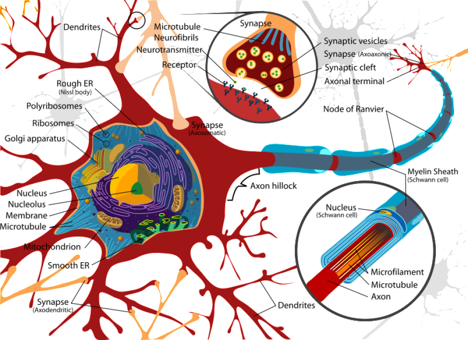
Brain Cell Drawing - Web 12 october 2023 this is the largest map of the human brain ever made researchers catalogue more than 3,000 different types of cell in our most complex organ. The nucleus of the neuron is found in the soma. The various parts of the brain are responsible for personality,. This is divided into three funiculi (anterior, lateral, and posterior) containing pathways travelling between the brain and the periphery. Web the second section shows how those many cells integrate to create sensory systems. The cerebrum (front of brain) comprises gray matter (the cerebral cortex) and white matter at its center. The largest part of the brain, the. Web anatomy explorer hindbrain and midbrain brain stem inferior colliculus medulla oblongata pons quadrigeminal lamina superior colliculus cerebellum cerebellar peduncle 4th ventricle cerebral aqueduct choroid plexus forebrain diencephalon choroid plexus of 3rd ventricle optic chiasm hypothalamic sulcus. Web contrary to the brain, the spinal cord’s outermost layer is formed of white matter. To isolate vascular cells from prenatal human brain tissues, we adapted our previously published protocol that was designed for the adult mouse brain 20,21.first, prenatal. Cajal, drawing from memory, has. The nucleus of the neuron is found in the soma. The two main types of cells in the brain are neurons, also known as. Given their diversity of functions performed in different parts of the nervous system, there is a wide variety in their shape, size, and electrochemical properties. Web there are two main types. Web cerebellum brainstem healthy brain summary the brain connects to the spine and is part of the central nervous system (cns). Web an easy guide to neuron anatomy with diagrams. Web anatomy explorer hindbrain and midbrain brain stem inferior colliculus medulla oblongata pons quadrigeminal lamina superior colliculus cerebellum cerebellar peduncle 4th ventricle cerebral aqueduct choroid plexus forebrain diencephalon choroid plexus. Web mini brains grown in a lab from stem cells spontaneously developed rudimentary eye structures, scientists reported in a fascinating paper in 2021. Web back in the 1890s, cajal produced a series of drawings of brain cells that would radically change scientists' understanding of the brain. This is divided into three funiculi (anterior, lateral, and posterior) containing pathways travelling between. Diagram of the components of a neuron. February 4, 2021 at 8:00 am. Neurons, also known as nerve cells, send and receive signals from your brain. Acting as a conduit, the axon carries these signals to. Web date december 13, 2023. Li and his colleagues published a paper in science entitled: The various parts of the brain are responsible for personality,. In the late 1800s, santiago ramón y cajal, a spanish brain scientist, spent long hours in his attic drawing elaborate cells. Neurons, also known as nerve cells, send and receive signals from your brain. Web contrary to the brain, the. Various processes (appendages or protrusions) extend from the cell body. By gemma conroy insights into. Web anatomy explorer hindbrain and midbrain brain stem inferior colliculus medulla oblongata pons quadrigeminal lamina superior colliculus cerebellum cerebellar peduncle 4th ventricle cerebral aqueduct choroid plexus forebrain diencephalon choroid plexus of 3rd ventricle optic chiasm hypothalamic sulcus. Web jane haley/cajal embroidery project. To isolate vascular. Li and his colleagues built up computational pipelines to analyze 1.1 million cells across 42 distinct human brain regions and identified 107 distinct cell types. Web back in the 1890s, cajal produced a series of drawings of brain cells that would radically change scientists' understanding of the brain. Neurons, also known as nerve cells, send and receive signals from your. Li and his colleagues built up computational pipelines to analyze 1.1 million cells across 42 distinct human brain regions and identified 107 distinct cell types. February 4, 2021 at 8:00 am. Neurons are highly specialized for the processing and transmission of cellular signals. Web anatomy explorer hindbrain and midbrain brain stem inferior colliculus medulla oblongata pons quadrigeminal lamina superior colliculus. Various processes (appendages or protrusions) extend from the cell body. To isolate vascular cells from prenatal human brain tissues, we adapted our previously published protocol that was designed for the adult mouse brain 20,21.first, prenatal. In the late 1800s, santiago ramón y cajal, a spanish brain scientist, spent long hours in his attic drawing elaborate cells. The two main types. Cajal, drawing from memory, has. By gemma conroy insights into. The rest of the brain tissue is structural or connective called the stroma which includes blood vessels. Web cell type isolation. Web the researchers analyzed more than 2.3 million individual brain cells from mice to create a comprehensive map of the mouse brain and used artificial intelligence to help predict. Web back in the 1890s, cajal produced a series of drawings of brain cells that would radically change scientists' understanding of the brain. Various processes (appendages or protrusions) extend from the cell body. Acting as a conduit, the axon carries these signals to. Web select a brain cells illustration for free download. Web 12 october 2023 this is the largest map of the human brain ever made researchers catalogue more than 3,000 different types of cell in our most complex organ. Neurons need to produce a lot of proteins, and most neuronal proteins are synthesized in the soma as well. Web anatomy explorer hindbrain and midbrain brain stem inferior colliculus medulla oblongata pons quadrigeminal lamina superior colliculus cerebellum cerebellar peduncle 4th ventricle cerebral aqueduct choroid plexus forebrain diencephalon choroid plexus of 3rd ventricle optic chiasm hypothalamic sulcus. The image shows an anatomical brain cross section, an abstraction of the brain with regions represented as colored circles (blue, red, green, and yellow), and a barcode to represent the technique used by the scientists. Neurons are highly specialized for the processing and transmission of cellular signals. Here, cajal’s images explore how the brain and sensory organs receive and process smells, sights and sounds. Web the second section shows how those many cells integrate to create sensory systems. Web cell type isolation. The two main types of cells in the brain are neurons, also known as. The rest of the brain tissue is structural or connective called the stroma which includes blood vessels. Amazing illustration images for your next project. Web salk institute researchers, as part of a larger collaboration with research teams around the world, analyzed more than half a million brain cells from three human brains to assemble an atlas of.
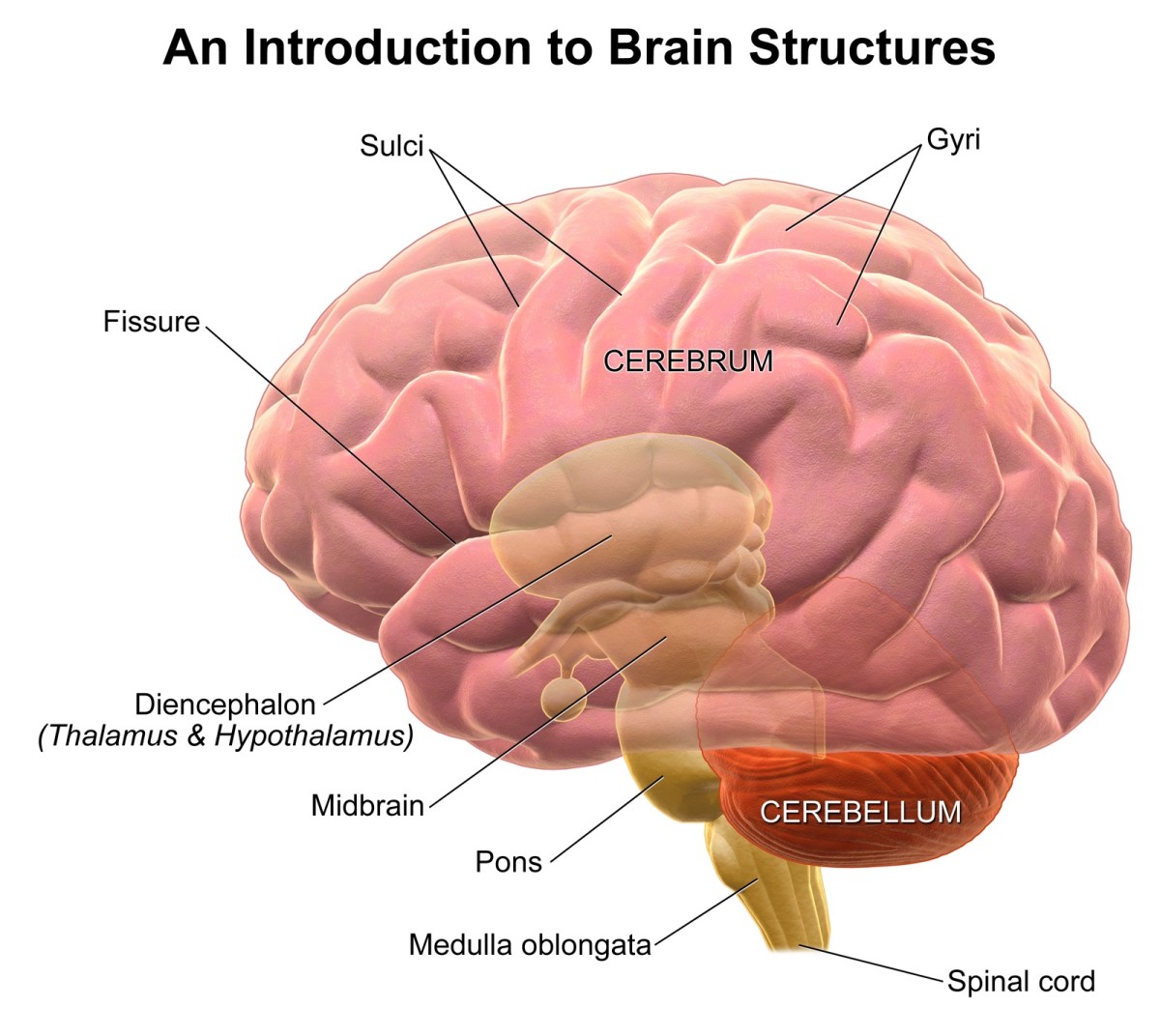
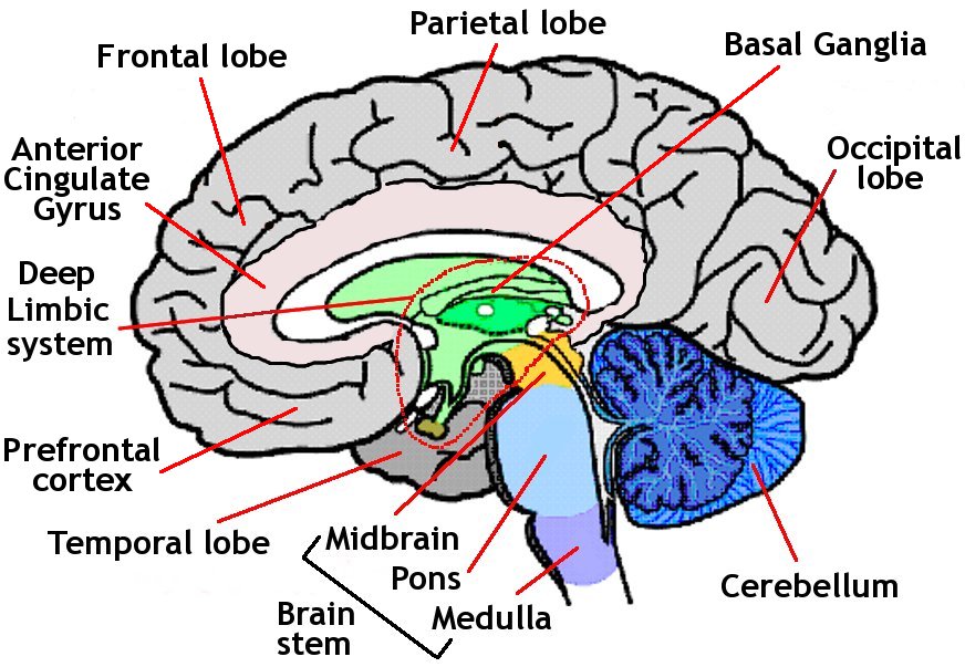
Brain 101 An Overview of the Anatomy and Physiology of the Brain

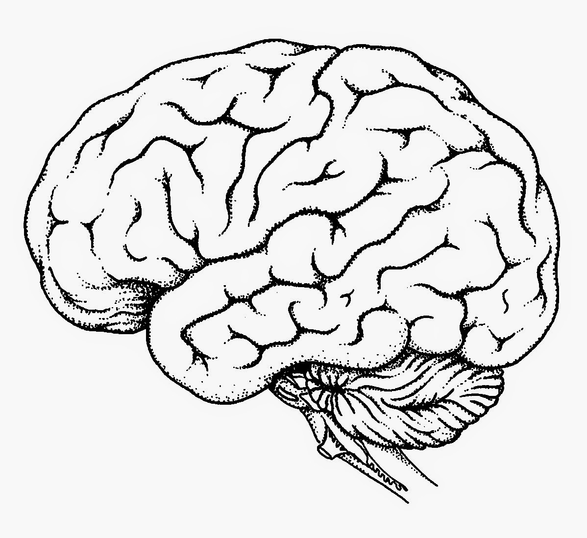
Human Brain Pencil Drawing bestpencildrawing

Brain cells, illustration Stock Image F013/1489 Science Photo Library

Brain Facts The Different Brain Cells
/human-nerve-cell--illustration-651425163-5b205b168e1b6e003681b4af.jpg)
The Brain Cells that can read Minds The Learning Zone

Brain Cell Medical, Health & Disease Pictures & Images Anatomy and

How Brain Cells Work Learnodo Newtonic

Human Brain Drawing at GetDrawings Free download

The Human Brain Facts, Anatomy, and Functions HubPages
.jpg)
Picture of human Brain Human Anatomy
A Group Of Scientists, Including Several At Harvard, Have Dived Deeper Into The Mammalian Brain Than Ever Before By Categorizing And Mapping At The Molecular Level All Of Its Thousands Of Different Cell Types.
Given Their Diversity Of Functions Performed In Different Parts Of The Nervous System, There Is A Wide Variety In Their Shape, Size, And Electrochemical Properties.
Cajal, Drawing From Memory, Has.
The Brain Has Been Called The Most Complex Structure In The Universe, But It May Also Be The Most Beautiful.
Related Post: