Cardiac Muscle Drawing
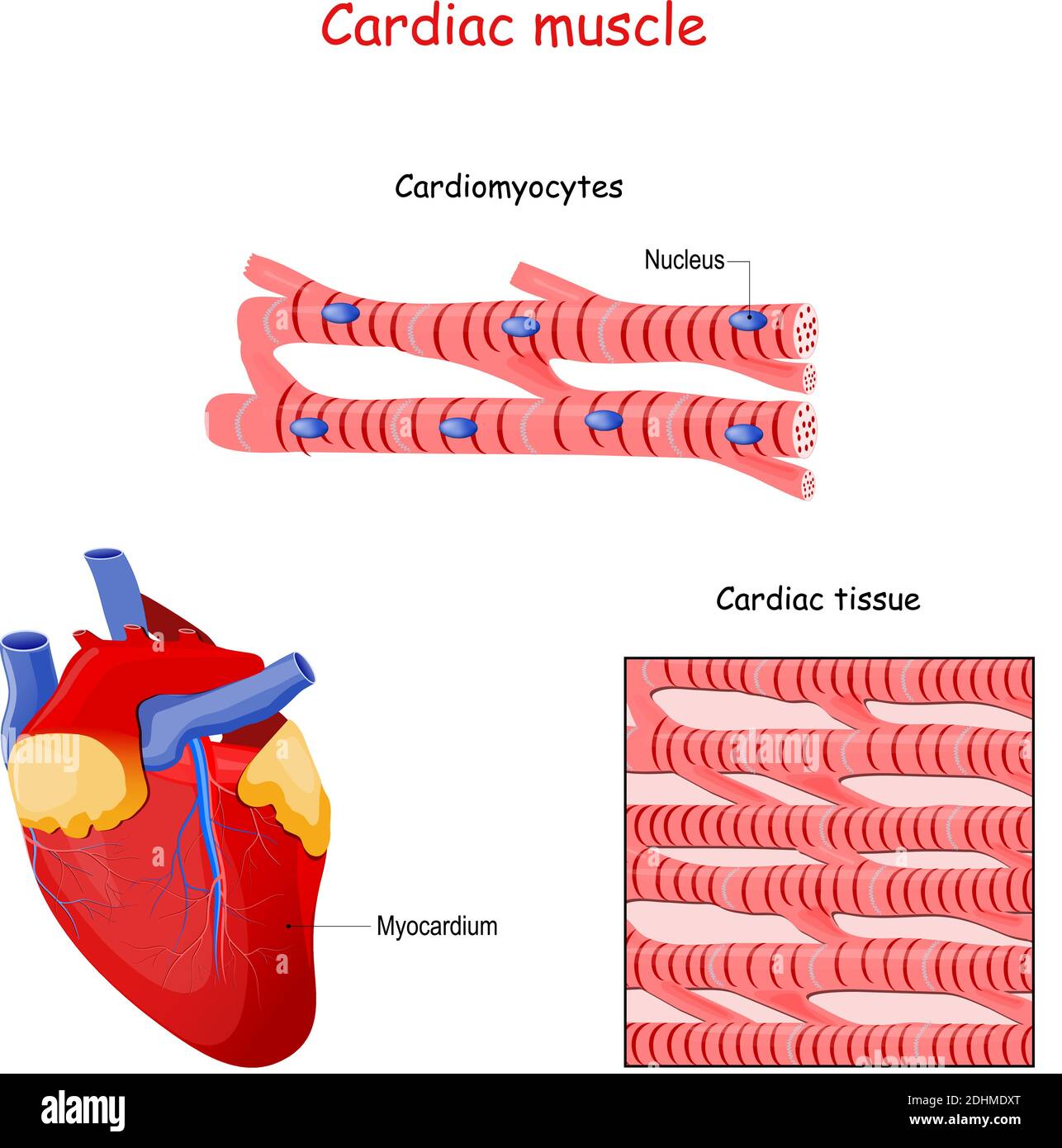
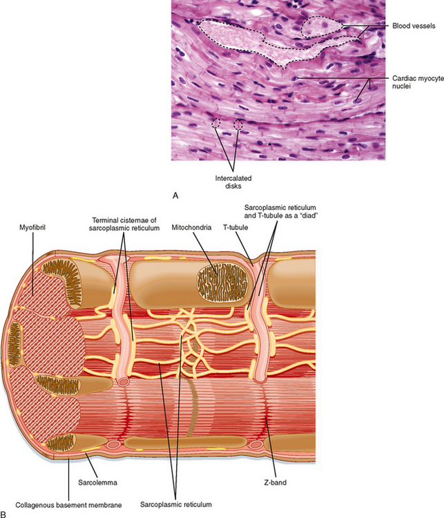
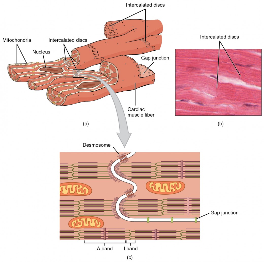
Cardiac Muscle Drawing - Cardiac muscle tissue is only found in the heart. The mitochondria in the cardiac myocytes are large and numerous. These inner and outer layers of the heart, respectively, surround the cardiac muscle tissue and separate it from the blood and other organs. Web by the end of this section, you will be able to: Diagrammatic view of three types. Web you will get the basic guide to learn cardiac muscle histology with real slide images and labeled diagrams. Web 1 2 sarcoplasmic reticulum tubules surround the myofibrils. Cardiac muscle tissue contracts and releases involuntarily. It is composed of elongated cells arranged in parallel that are capable of contracting and generating a force. Web 0:00 / 7:51 how to draw a muscle tissue| straight | smooth | cardiac fine arts guruji 438k subscribers 1.5k 80k views 2 years ago class 9 diagram how to draw a muscle tissue,straight. The mitochondria in the cardiac myocytes are large and numerous. Web let’s learn more about the cardiac muscle with the help of a diagram. Web how to draw cardiac muscle easily step by step | how to draw cardiac muscle 👉pencil colour diagrams? The myocardium is surrounded by a thin outer layer called the epicardium (aka visceral pericardium) and an. Myocardial contractile cells and myocardial conducting cells. Relate the structure of the heart to its function as a pump. Web cardiac muscle under microscope 40x, 100x, and 400x. Describe the structure of cardiac muscle. Cardiac muscle is made from sheets of cardiac muscle cells. Now, i will provide the cardiac muscle. Web cardiac muscle under microscope 40x, 100x, and 400x. Similar to skeletal muscle, cardiac muscle is striated and organized into sarcomeres, possessing the same banding organization as skeletal muscle ( [link] ). The human body quiz cardiac muscle cells form a highly branched cellular network in the heart. Web myocardial infarction cardiac hypertrophy. The myocardium is surrounded by a thin outer layer called the epicardium (aka visceral pericardium) and an inner endocardium. Web britannica quiz facts you should know: Web cardiac muscle (or myocardium) makes up the thick middle layer of the heart. Myocardial contractile cells and myocardial conducting cells. After the end of the article, i will share the cardiac muscle histology. Diagrammatic view of three types. It is composed of elongated cells arranged in parallel that are capable of contracting and generating a force. Compare the effect of ion movement on membrane potential of cardiac conductive and contractile cells. Web describe the internal and external anatomy of the heart. The mitochondria in the cardiac myocytes are large and numerous. Web cardiac muscle tissue, or myocardium, is a specialized type of muscle tissue that forms the heart. Cardiac muscle tissue is only found in the heart. Compare systemic circulation to pulmonary circulation. They are connected end to end by intercalated disks and are organized into layers of myocardial tissue that are wrapped around the chambers of the heart. Now, i. They are connected end to end by intercalated disks and are organized into layers of myocardial tissue that are wrapped around the chambers of the heart. Web in this video i have shown the simplest way of drawing muscle drawing. It performs involuntary, coordinated contractions that allow your heart to pump blood through. Web cardiac muscle also called the myocardium,. Web images structure cardiomyopathy exercise takeaway cardiac muscle tissue is only found in your heart. Myocardial contractile cells and myocardial conducting cells. Diagrammatic view of three types. Asapknowledge 31.1k subscribers subscribe 337 share 27k views 2 years ago #muscle #tissue #cardiac title: After the end of the article, i will share the cardiac muscle histology drawing with you. Web 0:00 / 7:51 how to draw a muscle tissue| straight | smooth | cardiac fine arts guruji 438k subscribers 1.5k 80k views 2 years ago class 9 diagram how to draw a muscle tissue,straight. Web cardiac muscle also called the myocardium, is one of three major categories of muscles found within the human body, along with smooth muscle and. Web cardiac muscle (or myocardium) makes up the thick middle layer of the heart. Contractile cells conduct impulses and are responsible for contractions that pump blood through the body. It performs involuntary, coordinated contractions that allow your heart to pump blood through. Web how to draw cardiac muscles step by step in a very easy way || type of muscles. Compare systemic circulation to pulmonary circulation. Web in this video you will learn how to draw cardiac muscle with easy step by step method.the individual cardiac muscle cell is a tubular structure composed of. Web how to draw diagram of cardiac muscle step by step for beginners ! Compare the effect of ion movement on membrane potential of cardiac conductive and contractile cells. Cardiac muscle tissue is only found in the heart. I will also enlist the functions and identification points of cardiac muscle. They are connected end to end by intercalated disks and are organized into layers of myocardial tissue that are wrapped around the chambers of the heart. It is one of three types of muscle in the body, along with skeletal and smooth muscle. Web 0:00 / 7:51 how to draw a muscle tissue| straight | smooth | cardiac fine arts guruji 438k subscribers 1.5k 80k views 2 years ago class 9 diagram how to draw a muscle tissue,straight. These inner and outer layers of the heart, respectively, surround the cardiac muscle tissue and separate it from the blood and other organs. The mitochondria in the cardiac myocytes are large and numerous. Web 1 2 sarcoplasmic reticulum tubules surround the myofibrils. Web images structure cardiomyopathy exercise takeaway cardiac muscle tissue is only found in your heart. Identify the veins and arteries of. Web myocardial infarction cardiac hypertrophy sources + show all muscle tissue muscle tissue is one of the four basic types of tissues that make up the human body. Web how to draw cardiac muscles step by step in a very easy way || type of muscles tissue hii, in this video , i will tell you that how can we draw the diag.
Cardiac Muscle Structure
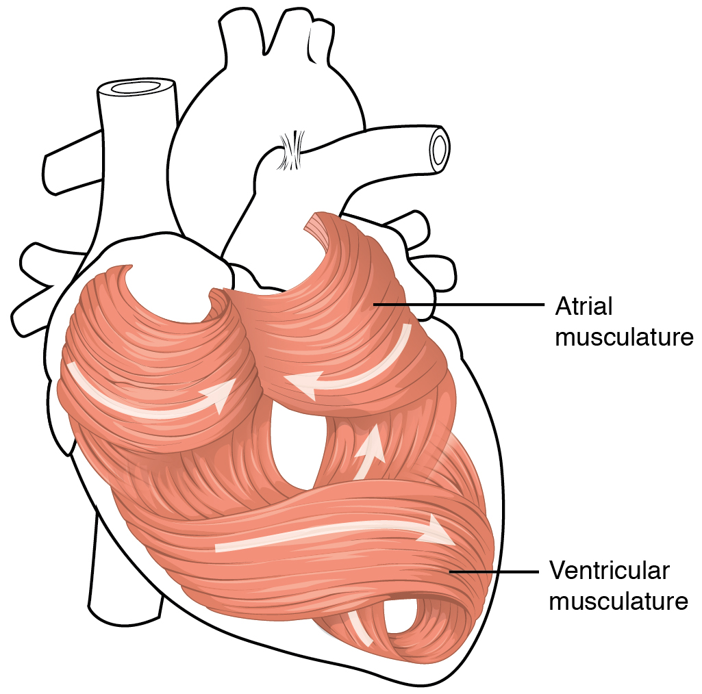
Heart Anatomy · Anatomy and Physiology
Cardiac Muscle Structure

Labeled Cardiac Muscle koibana.info Heart structure, Heart function
12.3 Types of Muscle Tissue Human Biology
Cardiac Muscle Basicmedical Key

Why are cardiac muscles so called? Explain their structure with the
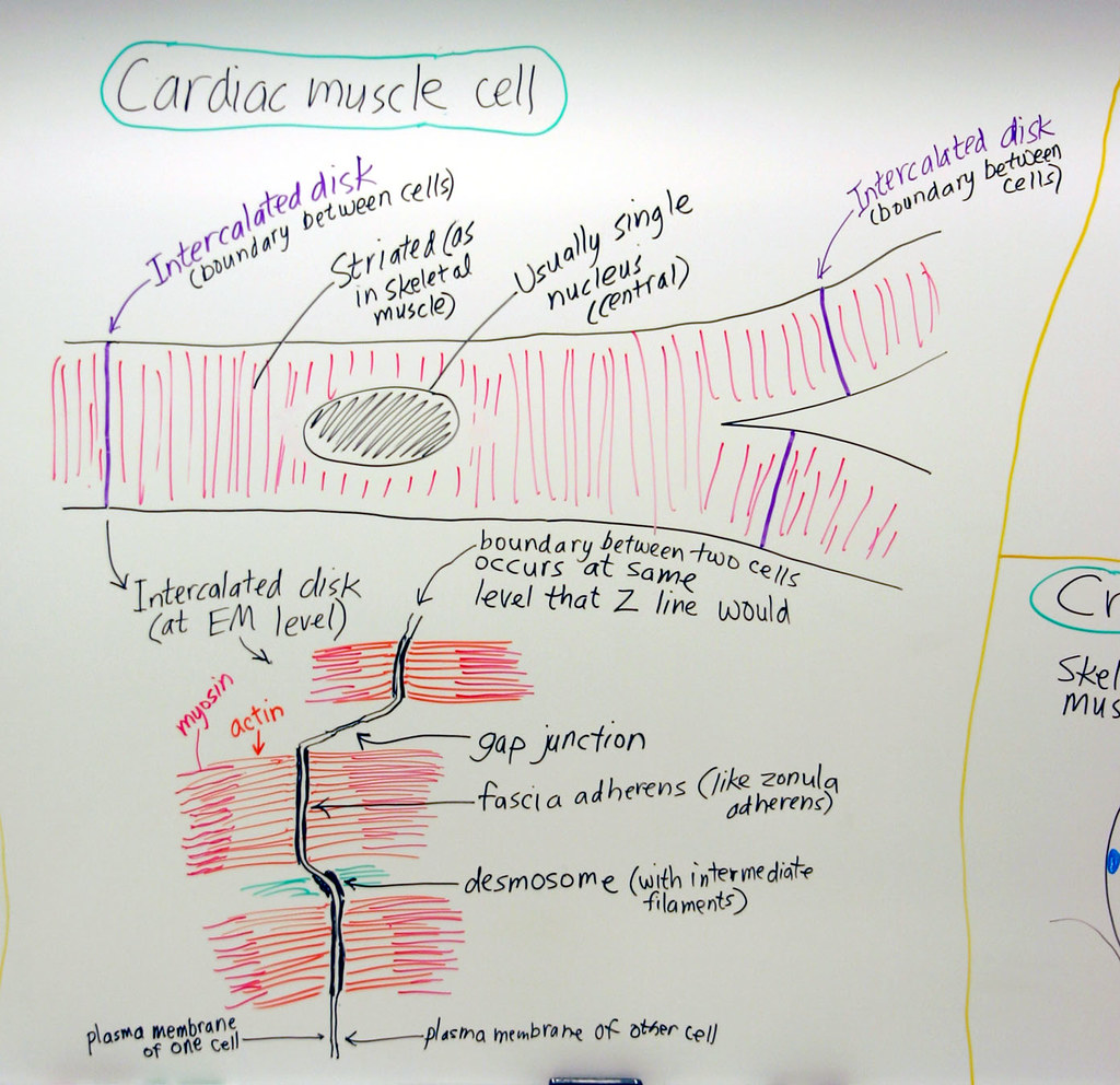
Muscle Cardiac Muscle Cell A hand drawn sketch by Dr. Chr… Flickr

How to draw " Cardiac Muscles" step by step in a very easy way Type
Cardiac Muscle and Electrical Activity Anatomy and Physiology II
Diagrammatic View Of Three Types.
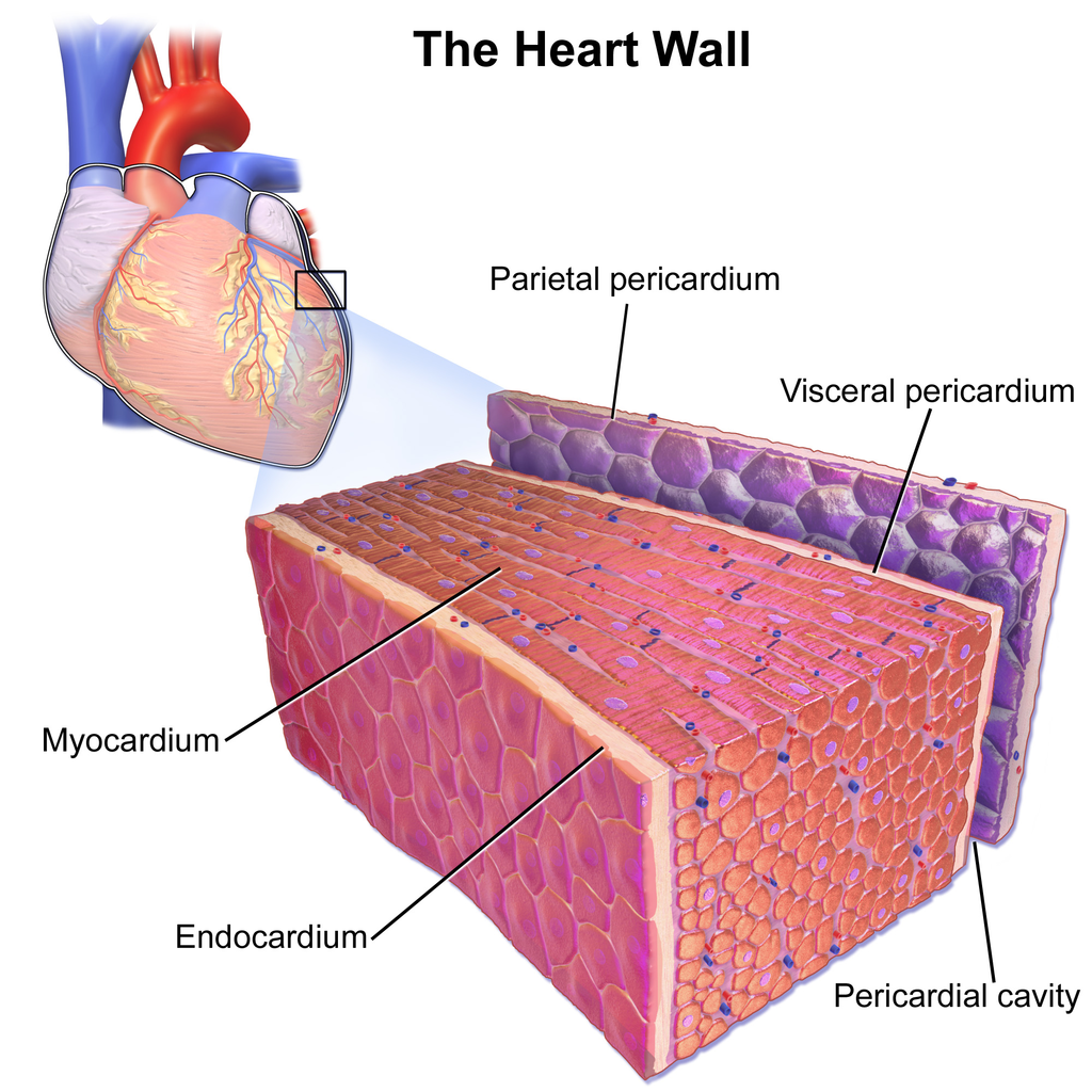
The Myocardium Is Surrounded By A Thin Outer Layer Called The Epicardium (Aka Visceral Pericardium) And An Inner Endocardium.
It Is The Pen Diagram Of Skeletal, Smooth And Cardiac Muscle For Class 10, 11 And 12.
Identify And Describe The Components Of The Conducting System That Distributes Electrical Impulses Through The Heart.
Related Post: