Draw And Label A Microscope
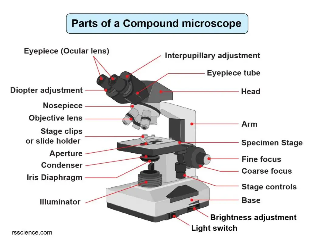
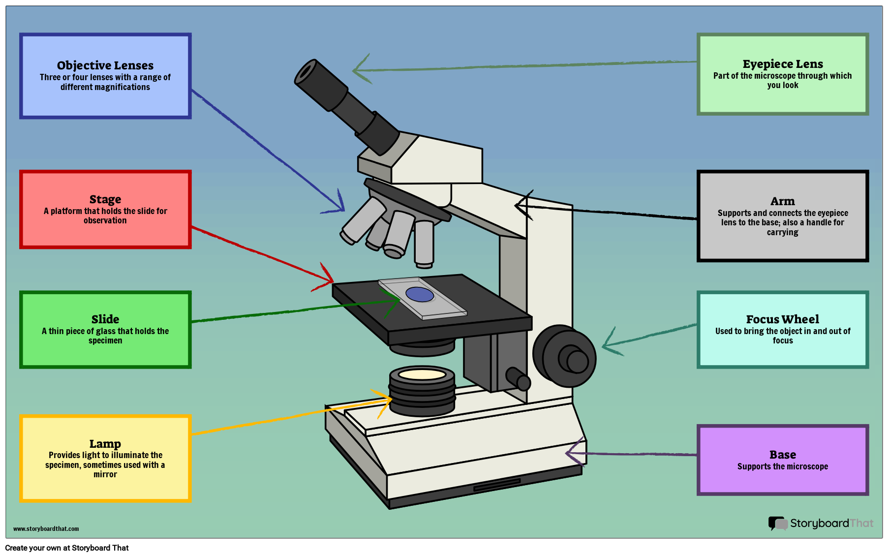
Draw And Label A Microscope - Web this activity has been designed for use in homes and schools. Begin with the eyepiece 1.2 step 2: Web a microscope is one of the invaluable tools in the laboratory setting. Web table of contents 1 how to draw a microscope that is hyperrealistic 1.1 step 1: Web in this interactive, you can label the different parts of a microscope. Outline the slide platform 1.6 step 6: Always lift a microscope by holding both the arm and base with two hands. Search for a diagram of a microscope. There are two major optical lens parts of a microscope: Shape the microscope head 1.3 step 3: Use a landscape poster layout (large or small). Always lift a microscope by holding both the arm and base with two hands. Web how to draw a microscope 🔬. Web there are three major structural parts of a microscope: However, as the saying goes, ‘practice makes perfect’, here is a blank compound microscope diagram. There are six printables available. First and foremost, we have a labeled microscope diagram, available in both black and white and color. Using arrows and textables label each part of the microscope and describe its function. Outline the slide platform 1.6 step 6: Mechanical parts of a compound microscope foot or base pillar arm stage inclination joint clips diaphragm nose. Kids will enjoy this simple step by step lesson for learning how to draw this essential piece of scientific equipment. The individual parts of a compound microscope can vary heavily depending on the configuration & applications that the scope is being used for. Also indicate the estimated cell size in micrometers under your drawing. Outline the slide platform 1.6 step. Web how to draw a microscope 🔬. Draw the objective lenses 1.5 step 5: Web these labeled microscope diagrams and the functions of its various parts, attempt to simplify the microscope for you. Young artists can learn to draw a microscope by following the photos in this easy guide. However, as the saying goes, ‘practice makes perfect’, here is a. It is also called a body tube or eyepiece tube. Web simple microscope is a magnification apparatus that uses a combination of double convex lens to form an enlarged, erect image of a specimen. Download the label the parts of the microscope pdf printable version here. Web a microscope is a laboratory instrument employed to examine minute entities, such as. Most photographs of cells are taken using a microscope, and these pictures can also be called micrographs. Learn how to use the microscope to view slides of several different cell types, including the use of the oil immersion lens to view bacterial cells. Knobs (fine and coarse) 6. To facilitate the scrutiny of such miniature entities with unparalleled. Using arrows. Useful as a means to change focus on one eyepiece so as to correct for any difference in vision between your two eyes. Learn how to use the microscope to view slides of several different cell types, including the use of the oil immersion lens to view bacterial cells. Web simple microscope is a magnification apparatus that uses a combination. First and foremost, we have a labeled microscope diagram, available in both black and white and color. Useful as a means to change focus on one eyepiece so as to correct for any difference in vision between your two eyes. Using arrows and textables label each part of the microscope and describe its function. Common compound microscope parts include: In. Download the label the parts of the microscope pdf printable version here. Stage and stage clips 7. Also indicate the estimated cell size in micrometers under your drawing. The working principle of a simple microscope is that when a lens is held close to the eye, a virtual, magnified and erect image of a specimen is formed at the least. There are six printables available. Young artists can learn to draw a microscope by following the photos in this easy guide. It is also called a body tube or eyepiece tube. Web ready to take your drawing skills to the next level? Always lift a microscope by holding both the arm and base with two hands. Web how to draw a microscope 🔬. Web simple microscope is a magnification apparatus that uses a combination of double convex lens to form an enlarged, erect image of a specimen. First and foremost, we have a labeled microscope diagram, available in both black and white and color. Draw the base of the microscope sketch 1.7 step 7: The working principle of a simple microscope is that when a lens is held close to the eye, a virtual, magnified and erect image of a specimen is formed at the least possible distance from which a human. Web there are three major structural parts of a microscope: The body tube connects the eyepiece to the objective lenses. Perfect for students or anyone. Web in this interactive, you can label the different parts of a microscope. Always lift a microscope by holding both the arm and base with two hands. Outline the arm frame 1.4 step 4: Web table of contents 1 how to draw a microscope that is hyperrealistic 1.1 step 1: There are three structural parts of the microscope i.e. Also indicate the estimated cell size in micrometers under your drawing. The scientific discipline encompassing the study of these diminutive entities through a microscope is called microscopy. Web each part of the compound microscope serves its own unique function, with each being important to the function of the scope as a whole.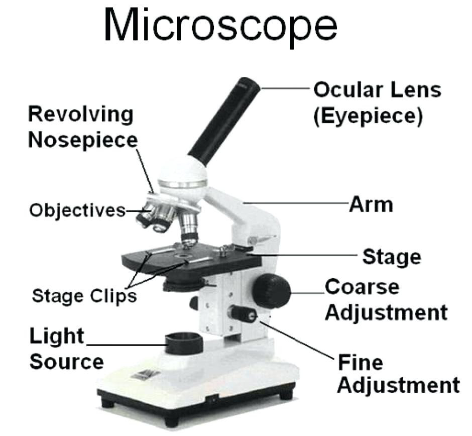
Parts Of A Microscope With Functions And Labeled Diagram Images

Parts of a microscope with functions and labeled diagram

Microscope Drawing And Label at GetDrawings Free download

Microscope Diagram Labeled, Unlabeled and Blank Parts of a Microscope
Compound Microscope Parts Labeled Diagram and their Functions (2023)

How to Use a Microscope (Properly) Step by Step New York Microscope
36+ Label Each Part Of A Microscope Gif Diagram Printabel

5 Types of Microscopes with Definitions, Principle, Uses, Labeled Diagrams

Microscope diagram Tom Butler Science skills, Microscope parts

Labeled Microscope Diagram Tim's Printables
Web This Activity Has Been Designed For Use In Homes And Schools.
Label The Cell Wall, Cell Membrane, Cytoplasm, And Chloroplasts In Your Lab Manual.
The Eyepiece Usually Contains A 10X Or 15X Power Lens.
Web Ready To Take Your Drawing Skills To The Next Level?
Related Post: