Occipital Bone Drawing
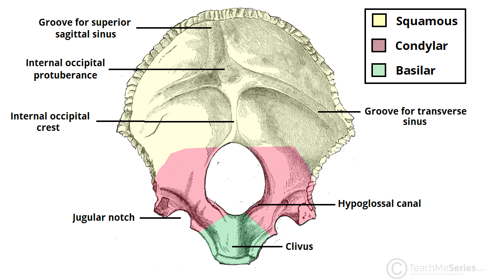
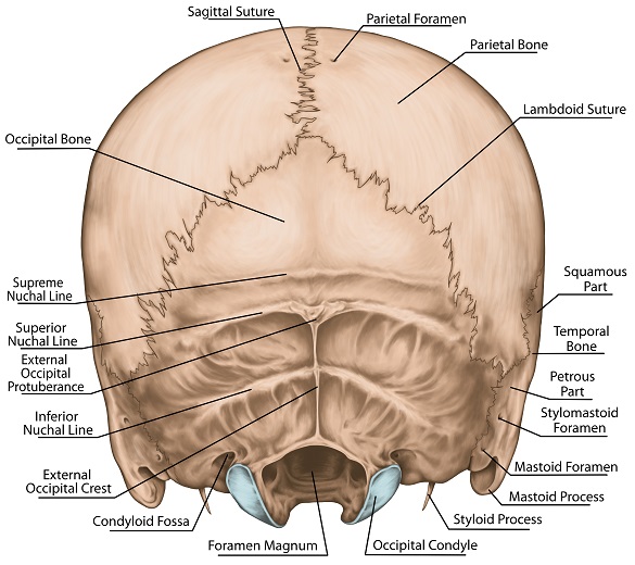
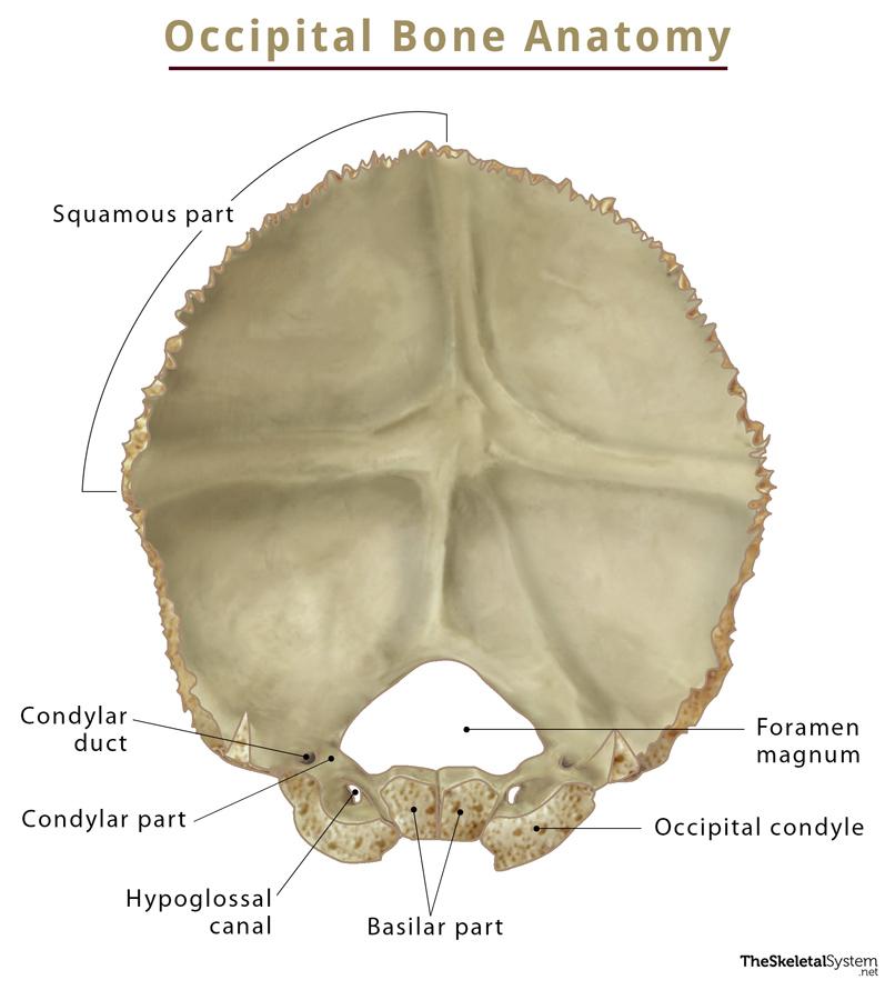
Occipital Bone Drawing - This article will describe the anatomy from the inferior view of the skull base. Web what is the occipital bone. Web the occipital bone is an unpaired bone which covers the back of the head (occiput). The occipital is an unpaired, trapezoidal cranial bone covering the back of the head. It is trapezoidal in shape. Jānis šavlovskis md, phd, assistant professor; Image retrieved from anatomy standard. External/internal surfaces basilar part (basiocciput): The occipital bone (os occipitale) is shown from superior. This image shows the different sulci created by the venous sinuses that pass along the internal surface of the os occipitale. Inferior view of the base of the skull [23:24] structures seen on the inferior view of the base of the skull. Web basics of drawing skulls when it comes to drawing skulls, there are a few things you need to keep in mind. It allows the spinal cord to pass from the brain into the spine. Image retrieved from anatomy. This article will describe the anatomy from the inferior view of the skull base. It has been improved and the format has been updated with more detailed examples. Lower/upper surfaces lateral (jugular) parts (two): This image shows the different sulci created by the venous sinuses that pass along the internal surface of the os occipitale. The external auditory canal adheres. The scalp, which consists of five layers, covers the bone. The occipital bone overlies the occipital lobes of the cerebrum. The sphenoid bone, petrous processes of the temporal bones, and the basilar part of the occipital bone. The internal surface of the squama (eminentia cruciformis, cruciform eminence) is caused by the cerebellum and cerebrum (resp. It is trapezoidal in shape. Image retrieved from anatomy standard. The occipital is cupped like a saucer in order to house the back part of the brain. Inferior view of the base of the skull [23:24] structures seen on the inferior view of the base of the skull. Web the occipital bone is the most posterior cranial bone and the main bone of the occiput.. The occipital bone (os occipitale) is shown from anterior. On its outside surface, at the posterior midline, is a small protrusion called the external occipital protuberance, which serves as an attachment site for a ligament of the posterior neck. So don’t try to copy someone else’s skull drawing; This image shows the different sulci created by the venous sinuses that. The occipital bone (os occipitale) is shown from posterior. The occipital is cupped like a saucer in order to house the back part of the brain. It is subdivided into the facial bones and the cranium, or cranial vault ( figure 7.3.1 ). The occipital bone (/ˌɒkˈsɪpɪtəl/) is a cranial dermal bone and the main bone of the occiput (back. The scalp, which consists of five layers, covers the bone. The occipital bone (os occipitale) is shown from posterior. The external auditory canal adheres closely to the bony surface of the temporal auditory canal. Web basics of drawing skulls when it comes to drawing skulls, there are a few things you need to keep in mind. The occipital bone (os. The occipital bone (os occipitale) is shown from posterior. The occipital is cupped like a saucer in order to house the back part of the brain. Web the occipital bone is the most posterior cranial bone and the main bone of the occiput. The occipital bone (os occipitale) is shown from anterior. Web what is the occipital bone. It contains outer and inner. It allows the spinal cord to pass from the brain into the spine. Image retrieved from anatomy standard. So don’t try to copy someone else’s skull drawing; Bodytomy explains the anatomy, diagram, and function of the occipital bone. The curved bone resembles a shallow dish. The zygomatic arches, mandibular fossae, tympanic plates and the styloid and mastoid processes. The occipital bone overlies the occipital lobe of the brain (which contains the primary. Image retrieved from anatomy standard. On its outside surface, at the posterior midline, is a small protrusion called the external occipital protuberance, which serves as an. Lower/upper surfaces lateral (jugular) parts (two): Where is the occipital bone located. The occipital bone is divided into four parts arranged around. It is trapezoidal in shape and curved on itself like a shallow dish. The sphenoid bone, petrous processes of the temporal bones, and the basilar part of the occipital bone. The occipital bone (os occipitale) is shown from anterolateral. External/internal surfaces basilar part (basiocciput): The scalp, which consists of five layers, covers the bone. Web basics of drawing skulls when it comes to drawing skulls, there are a few things you need to keep in mind. Bodytomy explains the anatomy, diagram, and function of the occipital bone. Web the occipital bone is the most posterior cranial bone and the main bone of the occiput. It allows the spinal cord to pass from the brain into the spine. This article will describe the anatomy from the inferior view of the skull base. The curved bone resembles a shallow dish. The external surface of the squamous part features: Instead, focus on capturing the essence of the skull and its individual features.Inner Surface of the Occipital Bone ClipArt ETC

The Occipital Bone Landmarks Attachments TeachMeAnatomy
Internal Surface of the Occipital Bone ClipArt ETC
Human Anatomy Scientific Illustrations Occipital Bone Stock

Occipital Bone ClipArt ETC

A Nod To The Occipital Bone And Your Health Body Wisdom CranioSacral

Occipital Bone The Definitive Guide Biology Dictionary

Occipital Bone The Definitive Guide Biology Dictionary

Occipital Bone Anatomy, Location, Functions, & Diagram

Occipital bone. Top view Anatomy bones, Occipital, Human anatomy
Consolidate Your Knowledge About The Base Of The Skull With The Following Quiz!
The Zygomatic Arches, Mandibular Fossae, Tympanic Plates And The Styloid And Mastoid Processes.
It Is Subdivided Into The Facial Bones And The Cranium, Or Cranial Vault ( Figure 7.3.1 ).
On Its Outside Surface, At The Posterior Midline, Is A Small Protrusion Called The External Occipital Protuberance, Which Serves As An Attachment Site For A Ligament Of The Posterior Neck.
Related Post:
