Parts Of A Microscope Drawing
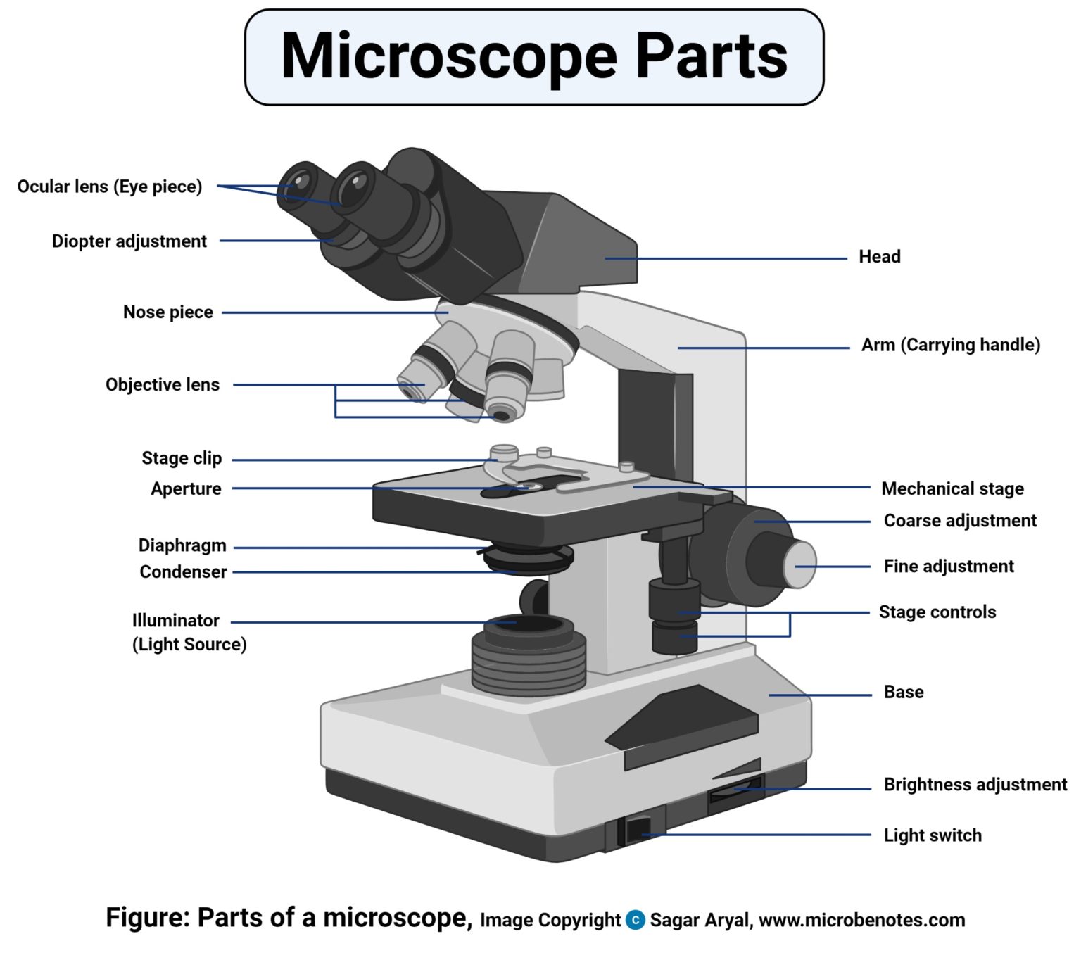
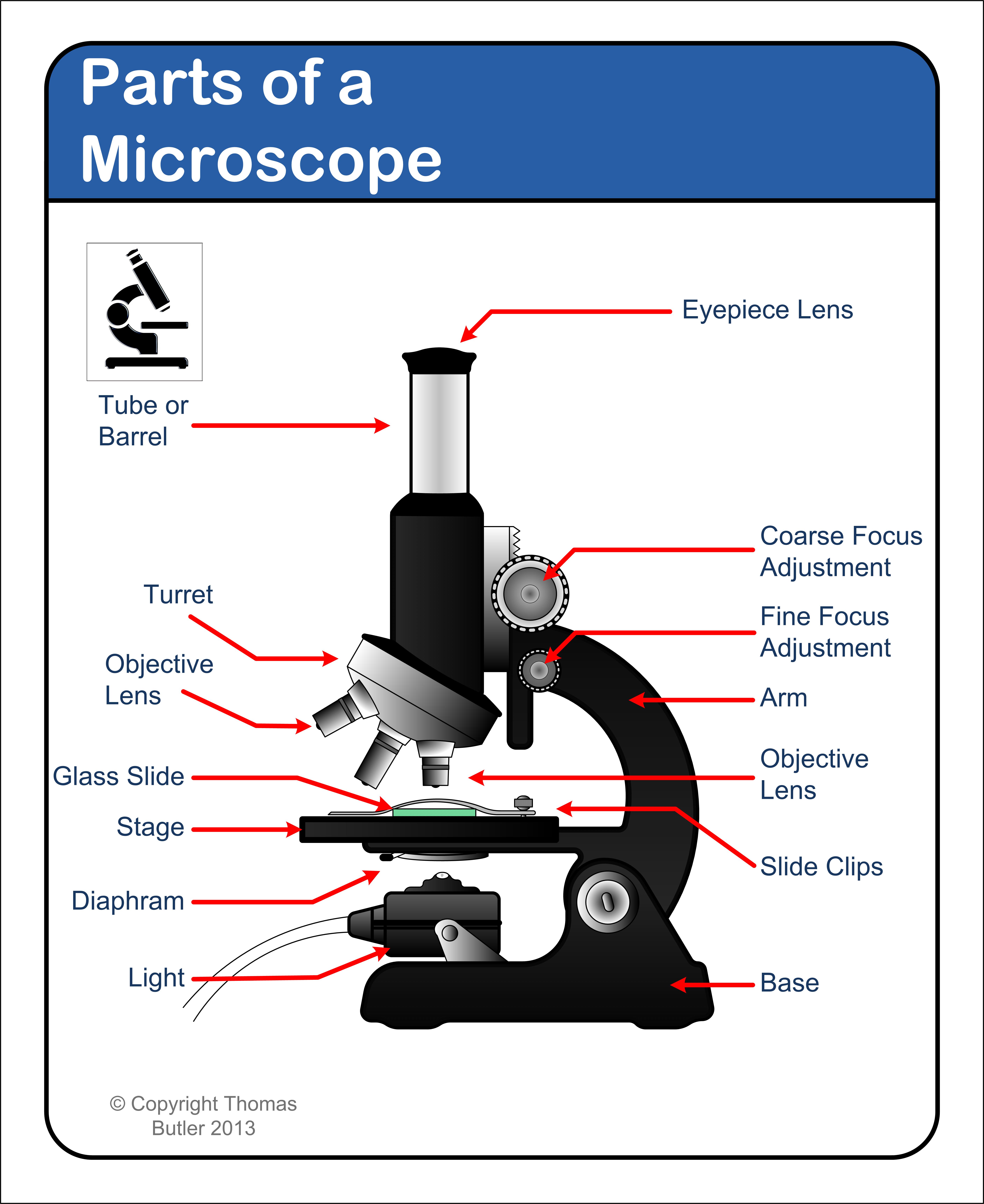
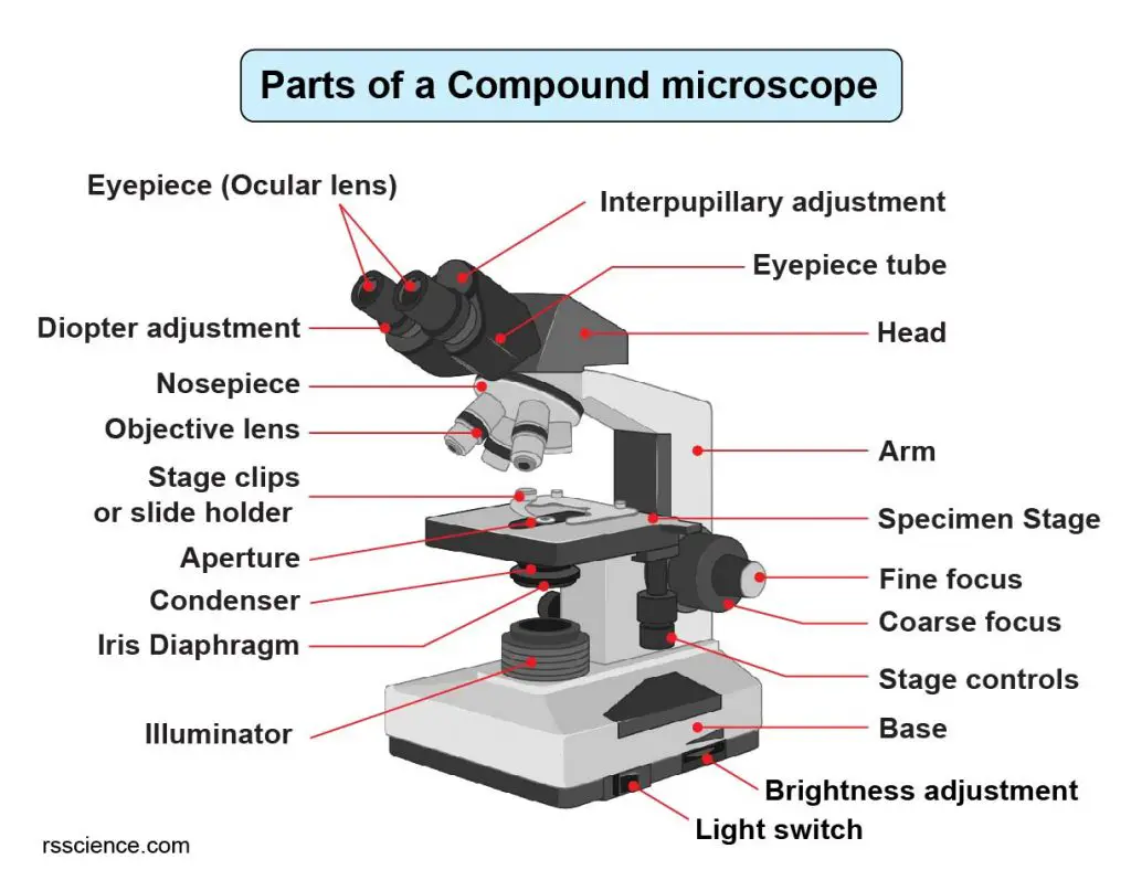
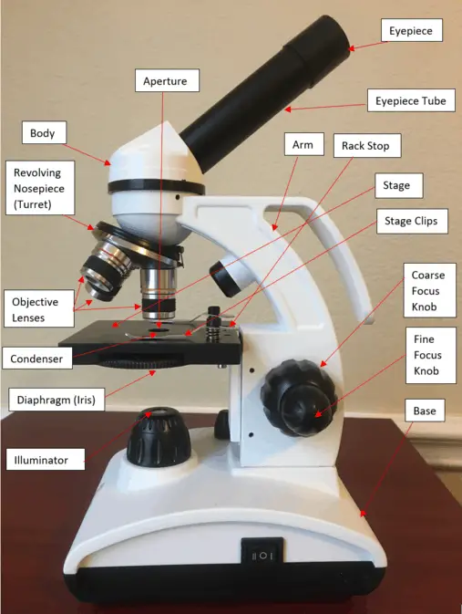
Parts Of A Microscope Drawing - The part that is looked through at the top of the compound. Shape the illuminator 1.8 step 8: The base acts as the foundation of microscopes and houses the illuminator. This type of microscope helps scientists look at tiny things like. Common compound microscope parts include: The head includes the upper part of the microscope, which houses the most critical optical components, and the eyepiece tube of the microscope. Web the 16 core parts of a compound microscope are: Web there are three major structural parts of a compound microscope. It is located at the top of the microscope, and the ocular lens or. Web exploring the compound microscope diagram. Web 1.1 step 1: The working principle of a simple microscope is that when a lens is held close to the eye, a virtual, magnified and erect image of a specimen is formed at the least possible distance from which a human. The arm connects between the base and the head parts. Web 📏 the microscope has three major structural. Draw the base of the microscope sketch 1.7 step 7: The arm connects between the base and the head parts. Outline the slide platform 1.6 step 6: Web learn about the microscope and its uses. A compound microscope is a special microscope with more than one lens. The head, the base, and the arm. Learn how to use the microscope to view slides of several different cell types, including the use of the oil immersion lens to view bacterial cells. Web simple microscope is a magnification apparatus that uses a combination of double convex lens to form an enlarged, erect image of a specimen. This type of. Web microscope parts labeled diagram; Carrie metzinger northover, bergmann lab, stanford university. Web let us take a look at the different parts of microscopes and their respective functions. Web the individual parts of a compound microscope can vary heavily depending on the configuration & applications that the scope is being used for. It is located at the top of the. The base acts as the foundation of microscopes and houses the illuminator. Begin with the eyepiece 1.2 step 2: Web 📏 the microscope has three major structural parts: Head (body) arm base eyepiece eyepiece tube objective lenses revolving nosepiece (turret) rack stop coarse adjustment knobs fine adjustment knobs stage stage clips aperture illuminator condenser diaphragm Web the resolution of a. Draw the objective lenses 1.5 step 5: Web microscope parts labeled diagram; Mechanical parts of a compound microscope foot or base pillar arm stage inclination joint clips diaphragm nose piece/revolving nosepiece/turret body tube adjustment knobs b. Resolution is expressed in linear units, usually micrometres (μm). Web simple microscope is a magnification apparatus that uses a combination of double convex lens. A compound microscope is a special microscope with more than one lens. The head includes the upper part of the microscope, which houses the most critical optical components, and the eyepiece tube of the microscope. Carrie metzinger northover, bergmann lab, stanford university. Web the individual parts of a compound microscope can vary heavily depending on the configuration & applications that. The part that is looked through at the top of the compound. Web today, we're learning how to draw a cool microscope!👩🎨 join our art hub membership! List down the three structural parts of a microscope. Web confocal microscopy image of a young leaf of thale cress, with one marker outlining the cells and other markers indicating young cells of. Through the eyepiece, you can visualize the object being studied. The head includes the upper part of the microscope, which houses the most critical optical components, and the eyepiece tube of the microscope. What are the mechanical parts? The most familiar type of microscope is the optical, or light, microscope, in which glass lenses are used to form the image.. Outline the slide platform 1.6 step 6: Web the 16 core parts of a compound microscope are: Web 📏 the microscope has three major structural parts: Learn about a microscopes parts and its functions including the eyepiece, objectives, and condenser with our labeled diagram. Mechanical parts pertain to the parts of the microscope that support the optional parts. Outline the slide platform 1.6 step 6: Head (body) arm base eyepiece eyepiece tube objective lenses revolving nosepiece (turret) rack stop coarse adjustment knobs fine adjustment knobs stage stage clips aperture illuminator condenser diaphragm Resolution is expressed in linear units, usually micrometres (μm). Brings the specimen into general focus. What are the mechanical parts? Web today, we're learning how to draw a cool microscope!👩🎨 join our art hub membership! Sometimes, people call it a biological microscope because scientists use it in labs. Web the resolution of a microscope is a measure of the smallest detail of the object that can be observed. Begin with the eyepiece 1.2 step 2: Web confocal microscopy image of a young leaf of thale cress, with one marker outlining the cells and other markers indicating young cells of the stomatal lineage (cells that will ultimately give rise to stomata, cellular valves used for gas exchange). Web parts of a compound microscope in its simplest form, the compound microscope consisted of two convex lenses aligned in series: The mechanical part and the optical parts. It has two main parts: A rotating turret that houses the objective lenses. Mechanical parts pertain to the parts of the microscope that support the optional parts. Coarse adjustment knobs bring the specimen into approximate focus, while fine adjustment knobs sharpen the focus quality of the image.
301 Moved Permanently

Microscope Diagram Labeled, Unlabeled and Blank Parts of a Microscope

Parts of a microscope with functions and labeled diagram

Diagrams of a Microscope 101 Diagrams

5 Types of Microscopes with Definitions, Principle, Uses, Labeled Diagrams

Simple Microscope Drawing at GetDrawings Free download

How to Use a Microscope

Compound Microscope Parts Labeled Diagram and their Functions Rs

Microscope Drawing And Label at GetDrawings Free download

16 Parts of a Compound Microscope Diagrams and Video Microscope Clarity
The Working Principle Of A Simple Microscope Is That When A Lens Is Held Close To The Eye, A Virtual, Magnified And Erect Image Of A Specimen Is Formed At The Least Possible Distance From Which A Human.
Explore The Different Parts Of A Microscope Using A Diagram, Including The Microscope Lens, Eyepiece, And Stage.
Draw The Objective Lenses 1.5 Step 5:
The Head, The Base, And The Arm.
Related Post: