Reticular Tissue Drawing
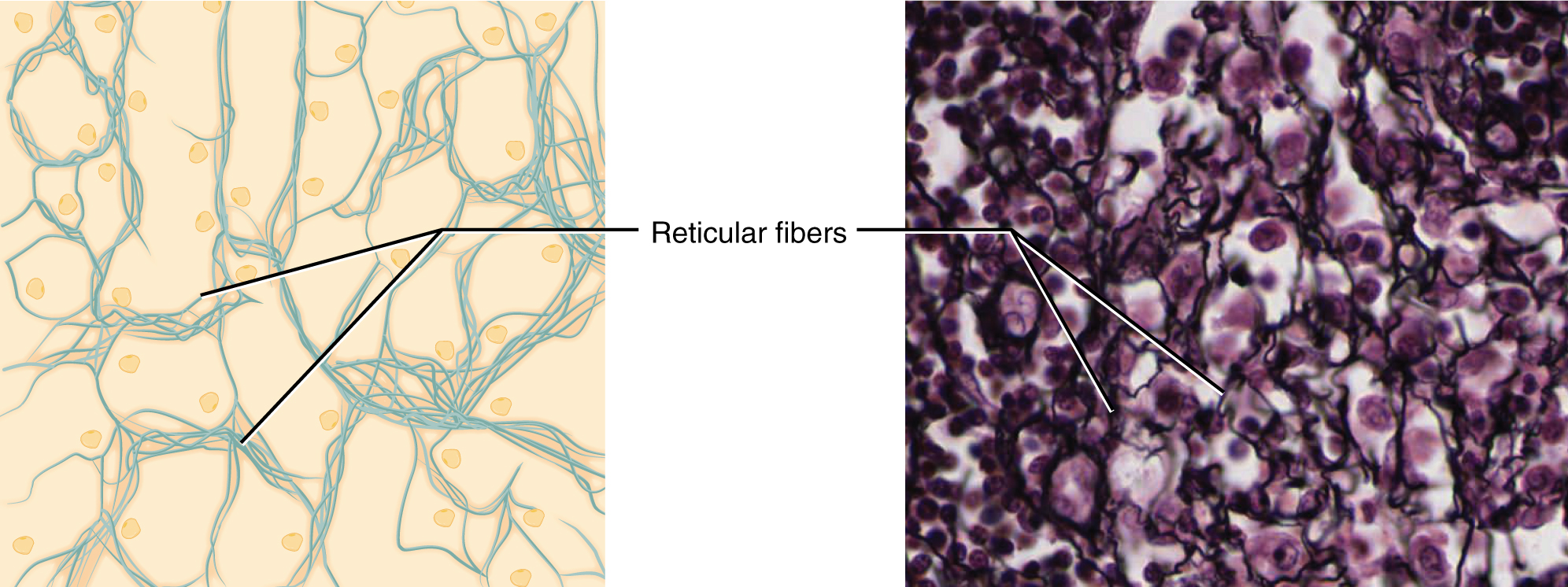
Reticular Tissue Drawing - It comprises a diverse group of cells that can be found in different parts of the body. None the human body has several types of tissues, a specific one being connective tissue. Web reticular tissue is a special subtype of connective tissue that is indistinguishable during routine histological staining. Please put total magnification in the image key. Fill out the blanks next to your drawing. The reticular fibers form the network onto which other cells attach. Web in 1858, at the university of erlangen in erlangen, germany, gerlach created a staining technique that yielded evidence to support his reticular theory. Reticular fibers are not unique to reticular connective tissue, but only in this type they are dominant. Web reticular tissue is a special subtype of connective tissue that is indistinguishable during routine histological staining. In the circle below, draw a representative sample of key features you identified, taking care to correctly and clearly draw their true shapes and directions. Loose connective tissue, adipose description (write or draw) draw an example. Web reticular connective tissue is named for the reticular fibers which are the main structural part of the tissue. Web reticular tissue is a type of connective tissue proper with an extracellular matrix consisting of an interwoven network of reticular fibers that provide a strong yet somewhat flexible framework. Reticular fibers are not unique to reticular connective tissue, but only in this type they are dominant. Web reticular connective tissue is named for the reticular fibers which are the main structural part of the tissue. Loose connective tissue textus connectivus laxus 1/3 synonyms: Use colored pencils and label if necessary. Web the most important function of this tissue is. In the circle below, draw a representative sample of key features you identified, taking care to correctly and clearly draw their true shapes and directions. Web reticular tissue is a special subtype of connective tissue that is indistinguishable during routine histological staining. • observe the characteristics of the three types of muscle Reticular meshes filter lymph and provide a microenvironment. Loose connective tissue, adipose description (write or draw) draw an example. Loose connective tissue textus connectivus laxus 1/3 synonyms: Loose connective tissue, reticular description (write or draw) draw an example. The specific types and relative proportions of cells, fibers, and ground substance determine the overall structure and function of connective tissues. Web reticular tissue is a special subtype of connective. Collagen fibers provide structure and tensile strength, with strands of collagen extending into both the papillary layer and the hypodermis. The cells that make the reticular fibers are fibroblasts called reticular cells. Web reticular connective tissue is a type of connective tissue [1] with a network of reticular fibers, made of type iii collagen [2] ( reticulum = net or. Reticular fibers form a dense structure, and hold together the cells of smooth muscle tissue, and also help in the formation of basement membrane. Please put total magnification in the image key. The cells that make the reticular fibers are fibroblasts called reticular cells. Web reticular tissue is a type of connective tissue proper with an extracellular matrix consisting of. Web connective tissue • comprises cells suspended in an extracellular matrix of protein fibers and ground substance. It comprises a diverse group of cells that can be found in different parts of the body. Loose connective tissue [10:11] structure and cellular components of loose connective tissue. Please put total magnification in the image key. Web obtain a slide of a. Elastin fibers provide some elasticity to the skin, enabling movement. • compare the interrelationship of epithelial and connective tissue through a study of the skin. Web connective tissue is a term used to describe a variety of types of tissues. Web reticular tissue supports the stroma of body organs, especially lymphoid. Loose connective tissue [10:11] structure and cellular components of. It is an important structural element that supports and separates spaces between our organs and tissues in the human body.the cells that make this connective tissue are loosely packed. Loose connective tissue, reticular description (write or draw) draw an example. Fill out the blanks next to your drawing. [3] reticular fibers are synthesized by special fibroblasts called reticular cells. Web. Web in 1858, at the university of erlangen in erlangen, germany, gerlach created a staining technique that yielded evidence to support his reticular theory. Loose connective tissue, adipose description (write or draw) draw an example. Reticular fibers form a dense structure, and hold together the cells of smooth muscle tissue, and also help in the formation of basement membrane. Web. Web in 1858, at the university of erlangen in erlangen, germany, gerlach created a staining technique that yielded evidence to support his reticular theory. Loose connective tissue [10:11] structure and cellular components of loose connective tissue. Web reticular tissue, a type of loose connective tissue in which reticular fibers are the most prominent fibrous component, forms the supporting framework of the lymphoid organs (lymph nodes, spleen, tonsils), bone marrow and liver. Reticular fibers are not unique to reticular connective tissue, but only in this type they are dominant. • observe the characteristics of the three types of muscle Reticular connective tissue forms a scaffolding for other cells in several organs, such as lymph nodes and bone marrow. Its subunits, the reticular fibers, are predominant structures in the human body, but they are mainly scattered and mixed with other types of fibers. • study the characteristics of loose, dense, elastic, and reticular connective tissue, adipose tissue, cartilage, and bone. Web reticular connective tissue 10x. It functions like a scaffolding that gives shape to these organs while also allowing space for blood and cells to pass through it. Web reticular connective tissue is named for the reticular fibers which are the main structural part of the tissue. Use colored pencils and label if necessary. Collagen fibers provide structure and tensile strength, with strands of collagen extending into both the papillary layer and the hypodermis. Web connective tissue is a term used to describe a variety of types of tissues. None the human body has several types of tissues, a specific one being connective tissue. The specific types and relative proportions of cells, fibers, and ground substance determine the overall structure and function of connective tissues.
Reticular Connective Tissue, 40X Histology

Reticular Connective Tissue

Reticular Connective Tissue 20x Histology

Reticular connective Tissue Diagram Quizlet

Reticular connective tissue cells and structure (preview) Human

Reticular Connective Tissue Diagram Quizlet

Reticular connective tissue Microscopic cells, Loose connective

Reticular Connective Tissue

Reticular Connective Tissue Structure
Connective Tissue Supports and Protects · Anatomy and Physiology
It Comprises A Diverse Group Of Cells That Can Be Found In Different Parts Of The Body.
Reticular Meshes Filter Lymph And Provide A Microenvironment For The Passage And Attachment Of White Blood Cells.
Reticular Fibers Form A Dense Structure, And Hold Together The Cells Of Smooth Muscle Tissue, And Also Help In The Formation Of Basement Membrane.
Loose Connective Tissue Textus Connectivus Laxus 1/3 Synonyms:
Related Post: