Skeletal Muscle Drawing
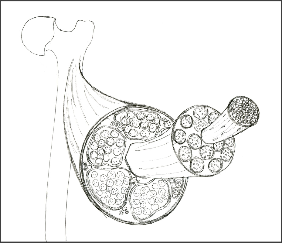
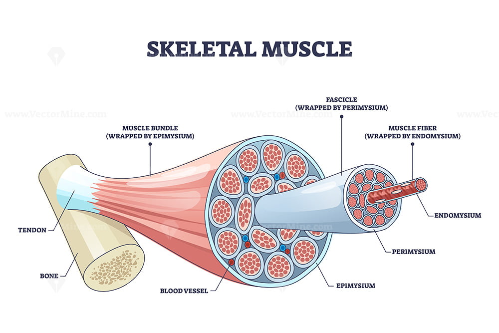
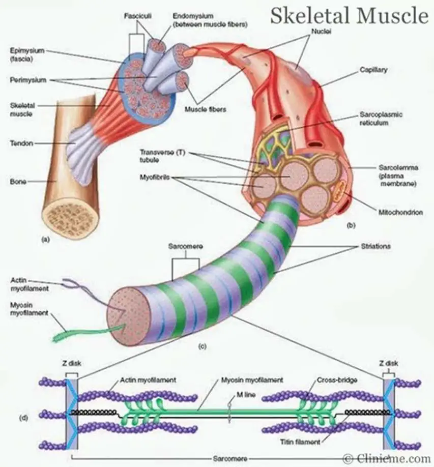
Skeletal Muscle Drawing - Identifying features are cylindrical cells and multiple peripheral nuclei. Each organ or muscle consists of skeletal muscle tissue, connective tissue, nerve tissue, and blood or vascular tissue. #muscle #diagram #howtodrawthis is a drawing of structure of the skeletal muscle. Several muscle fibers gathered together into a bundle are called a muscle fascicle. These tissues include the skeletal muscle fibers, blood vessels, nerve fibers, and connective tissue. Web i think we have a respectable sense of how muscles contract on the molecular level. Skeletal muscles, in particular, are the ones that act on the body joints to produce movements. These muscle cells are slender and long and are termed as muscle fibres. Within muscles, there are layers of connective tissue called the epimysium, perimysium, and endomysium. Diagrammatic view of three types. Skeletal muscles vary considerably in size, shape, and arrangement of fibers. Each organ or muscle consists of skeletal muscle tissue, connective tissue, nerve tissue, and blood or vascular tissue. Identifying features are cylindrical cells and multiple peripheral nuclei. There are three main types of muscle: Web i think we have a respectable sense of how muscles contract on the molecular. Web hello friends, this is my youtube channel and in this channel i used to share videos of different diagrams in easy way and step by step tutorials. That's their elbow and let's say that's their hand right there. Use the bony landmarks prominent bones will help you navigate the skeletal structure to help identify the placement. Web anatomy of. Longitudinal section of skeletal muscle #2. Skeletal muscles vary considerably in size, shape, and arrangement of fibers. A whole skeletal muscle is considered an organ of the muscular system. Drawing of a skeletal muscle stock illustrations. Web the musculoskeletal system (locomotor system) is a human body system that provides our body with movement, stability, shape, and support. Skeletal muscles act not only to produce movement but also to stop movement, such as resisting gravity to maintain posture. Muscular system, which includes all types of muscles in the body. Web hello friends, this is my youtube channel and in this channel i used to share videos of different diagrams in easy way and step by step tutorials. Web. Within muscles, there are layers of connective tissue called the epimysium, perimysium, and endomysium. Each fascicle is wrapped in a collagen sheath, called a perimysium. If you don't have a printer just keep this open. Muscle tissue has a unique histological appearance which enables it to carry out its function. A whole skeletal muscle is considered an organ of the. Skeletal muscles vary considerably in size, shape, and arrangement of fibers. It is subdivided into two broad systems: Learn step by step drawing tutorial. Use the bony landmarks prominent bones will help you navigate the skeletal structure to help identify the placement. Muscular system, which includes all types of muscles in the body. It is the pen diagram of skeletal, smooth and cardiac muscle for class 10, 11 and 12. These tissues include the skeletal muscle fibers, blood vessels, nerve fibers, and connective tissue. There are three main types of muscle: Skeletal muscle injuries or diseases can have a profound effect on your life. Download a free printable outline of this video and. There are three main types of muscle: Web every skeletal muscle in your body is made up of hundreds of thousands of tiny muscle fibers (or muscle cells). Skeletal muscles vary considerably in size, shape, and arrangement of fibers. Extensible tissue can be stretched and elastic tissue is able to return to its original shape following distortion. Longitudinal section of. Extensible tissue can be stretched and elastic tissue is able to return to its original shape following distortion. These tissues include the skeletal muscle fibers, blood vessels, nerve fibers, and connective tissue. It is subdivided into two broad systems: Each muscle fiber is wrapped in a connective tissue sheath, called an endomysium. Web ultrastructure of muscle cells. View, isolate, and learn human anatomy structures with zygote body. These layers cover muscle subunits, individual muscle cells, and myofibrils respectively. It is subdivided into two broad systems: Several muscle fibers gathered together into a bundle are called a muscle fascicle. Skeletal muscles vary considerably in size, shape, and arrangement of fibers. Skeletal muscle injuries or diseases can have a profound effect on your life. Web anatomy of a skeletal muscle cell. Web every skeletal muscle in your body is made up of hundreds of thousands of tiny muscle fibers (or muscle cells). Download a free printable outline of this video and draw along with us. Longitudinal section of skeletal muscle #2. Skeletal muscles act not only to produce movement but also to stop movement, such as resisting gravity to maintain posture. Diagrammatic view of three types. There are three layers of connective tissue: Learn step by step drawing tutorial. Skeletal muscle fibers of the longitudinal section #3. Let's take a step back now and just understand how muscles look, at least structurally, or how they relate to things that we normally associate with muscles. Muscles work on a macro level, starting with tendons that attach muscles to bones. Web in this video i have shown the simplest way of drawing muscle drawing. Identifying features are cylindrical cells and multiple peripheral nuclei. So let me draw a flexing bicep right here. Several muscle fibers gathered together into a bundle are called a muscle fascicle.
Skeletal Muscle Structure Amanda Barnaby

Skeletal muscle, illustration Stock Image C006/3938 Science Photo

Skeletal muscle structure with anatomical inner layers outline diagram

(A) Illustration of skeletal muscle structure copied with permission

How To Draw Structure Of Skeletal Muscle YouTube

Schematic representation of the skeletal muscle structure. The

Skeletal muscle diagram Healthiack

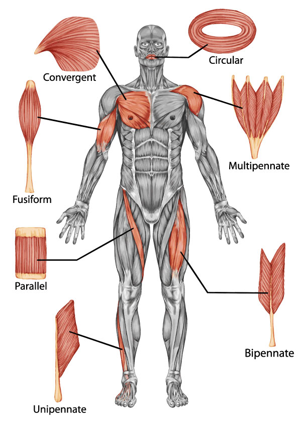
Skeletal Muscle Shapes

Shapes of skeletal muscles with various muscular types outline diagram

Introduction to Skeletal Muscle Boundless Anatomy and Physiology
Each Organ Or Muscle Consists Of Skeletal Muscle Tissue, Connective Tissue, Nerve Tissue, And Blood Or Vascular Tissue.
Web How To Draw Human Muscular System.
Blood Vessels And Nerves Enter The Connective Tissue And Branch In The Cell.
Muscle Tissue Has A Unique Histological Appearance Which Enables It To Carry Out Its Function.
Related Post: