Smooth Muscle Tissue Drawing
Smooth Muscle Tissue Drawing - Unlike cardiac and skeletal muscle cells, smooth muscle cells do not exhibit striations since their actin and myosin (thin and thick) protein filaments are not organized as sarcomeres. Web smooth muscle can be confused with cardiac muscle because the cells are often running in different directions, just as they are in cardiac muscle. Web smooth muscles are widely distributed in the animal’s body and predominantly found in the visceral hollow organs and blood vessels. The area inside the box is enlarged in the next image. In the circle below, draw a representative sample of key features you identified, taking care to correctly and clearly draw their true shapes and directions. Smooth muscle fibers are often found forming sheets of tissue and function in a coordinated fashion due to the presence of gap junctions between the cells. This is because smooth muscle contributes to many different tissues throughout the body. Web smooth muscle is a type of muscle tissue which is used by various systems to apply pressure to vessels and organs. Web how to draw a muscle tissues/how to draw striated smooth and cardiac muscles.it is very easy drawing detailed method to help you.i draw the cardiac muscles. It is the pen diagram of skeletal, smooth and cardiac muscle for class 10, 11 and 12. This is because smooth muscle contributes to many different tissues throughout the body. Smooth muscle cells are a lot smaller than cardiac muscle cells, and they do not branch or connect end to end the way cardiac cells do. Also, smooth muscle tissue is mostly cellular (and therefore more nuclei are present), whereas the connective tissue is mostly extracellular collagen. Web smooth muscle is a type of muscle tissue which is used by various systems to apply pressure to vessels and organs. The area inside the box is enlarged in the next image. Fill out the blanks next to your drawing. These cells have fibers of actin and myosin which run through the cell and are supported by a framework. Web obtain a slide of smooth muscle tissue from the slide box. Web author information and affiliations last update: This is because smooth muscle contributes to many different tissues throughout the body. It is in the stomach and intestines where it. The table below compares the differences in the morphology of the three types of. Also, smooth muscle tissue is mostly cellular (and therefore more nuclei are present), whereas the connective tissue is mostly extracellular collagen fibers with fewer cells. Smooth muscle cells are a lot smaller than cardiac muscle cells, and they do not branch or connect end to end the way cardiac cells do. Web smooth muscle can be confused with cardiac muscle. This is because smooth muscle contributes to many different tissues throughout the body. Web smooth muscle is a type of muscle tissue which is used by various systems to apply pressure to vessels and organs. In the circle below, draw a representative sample of key features you identified, taking care to correctly and clearly draw their true shapes and directions.. Smooth muscle cells are a lot smaller than cardiac muscle cells, and they do not branch or connect end to end the way cardiac cells do. Web smooth muscle is one of three types of muscle tissue, alongside cardiac and skeletal muscle. Web smooth muscles are widely distributed in the animal’s body and predominantly found in the visceral hollow organs. Web smooth muscle is a type of tissue found in the walls of hollow organs, such as the intestines, uterus and stomach. The area inside the box is enlarged in the next image. Smooth muscle is composed of sheets or strands of smooth muscle cells. This is because smooth muscle contributes to many different tissues throughout the body. Web smooth. These cells have fibers of actin and myosin which run through the cell and are supported by a framework of other proteins. One unique feature of neural crest cells is that their migration occurs. Web author information and affiliations last update: They range from about 30 to 200 μ m (thousands of times shorter than skeletal muscle fibers), and they. Web smooth muscle is found throughout the body around various organs and tracts. Web smooth muscle derives from both mesoderm and neural crest cells; This is because smooth muscle contributes to many different tissues throughout the body. The table below compares the differences in the morphology of the three types of. Web in this video i have shown the simplest. Web smooth muscle is a type of tissue found in the walls of hollow organs, such as the intestines, uterus and stomach. It is in the stomach and intestines where it. Web smooth muscle derives from both mesoderm and neural crest cells; Web smooth muscle is found throughout the body around various organs and tracts. Web smooth muscle is one. This is because smooth muscle contributes to many different tissues throughout the body. Also, smooth muscle tissue is mostly cellular (and therefore more nuclei are present), whereas the connective tissue is mostly extracellular collagen fibers with fewer cells. It is the pen diagram of skeletal, smooth and cardiac muscle for class 10, 11 and 12. Individual cells range in size from 30 to 200 μm. They range from about 30 to 200 μ m (thousands of times shorter than skeletal muscle fibers), and they produce their own connective tissue, endomysium. In the circle below, draw a representative sample of key features you identified, taking care to correctly and clearly draw their true shapes and directions. Smooth muscle is composed of sheets or strands of smooth muscle cells. You can also find smooth muscle in the walls of passageways , including arteries and veins of de cardiovascular system. How to draw a muscle tissue,straight muscles,smooth muscles,cardiac muscles ══🅒🅛🅐🅢🅢 11th. Web smooth muscle derives from both mesoderm and neural crest cells; The area inside the box is enlarged in the next image. Web author information and affiliations last update: Smooth muscle cells can undergo hyperplasia, mitotically dividing to produce new cells. Unlike cardiac and skeletal muscle cells, smooth muscle cells do not exhibit striations since their actin and myosin (thin and thick) protein filaments are not organized as sarcomeres. One unique feature of neural crest cells is that their migration occurs. Web in this video i have shown the simplest way of drawing muscle drawing.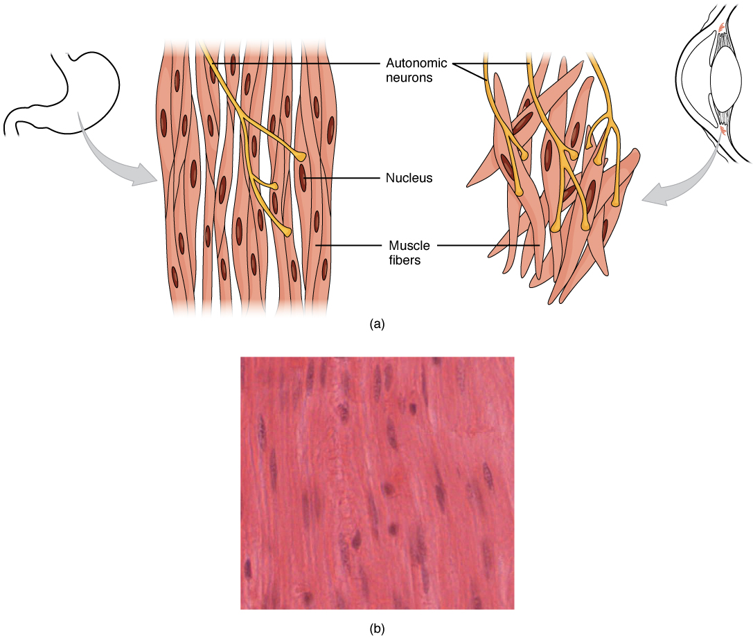
Smooth Muscle Anatomy and Physiology I

How To Draw Muscle Fibers at How To Draw
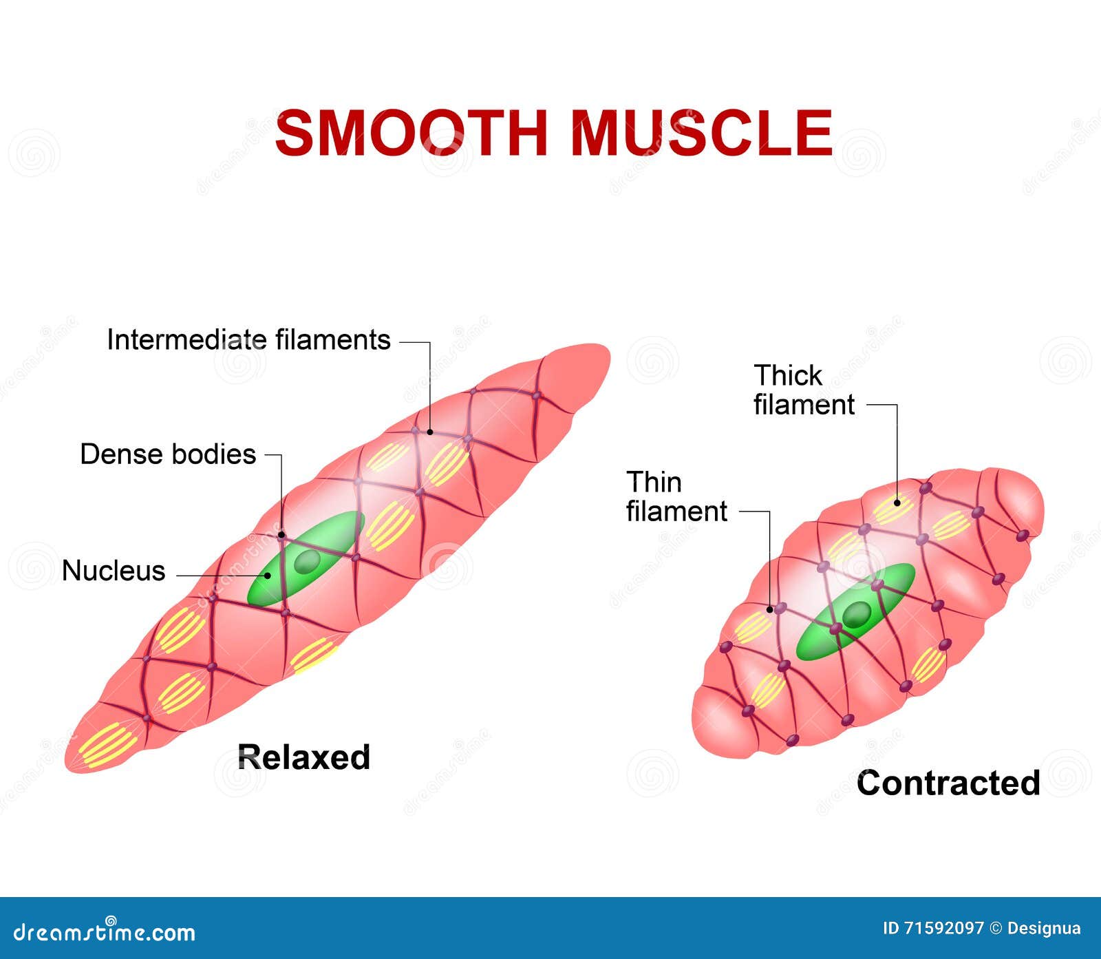
Smooth muscle tissue stock vector. Illustration of autonomic 71592097

Smooth Muscle Diagram Smooth Muscle Examples And Function
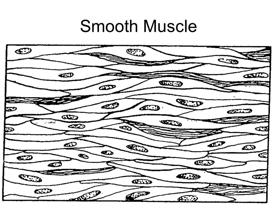
Smooth Muscle Diagram Drawing Smooth Muscle Structure Function
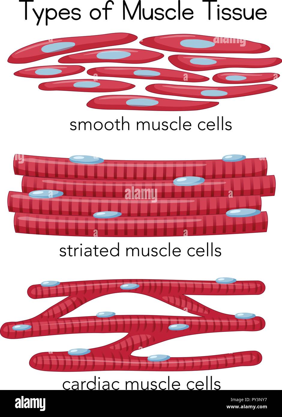
Types of Muscle Tissue illustration Stock Vector Image & Art Alamy
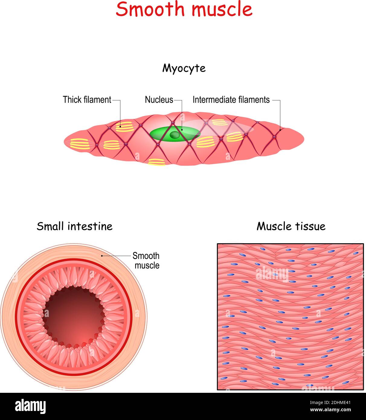
Structure of smooth muscle fibers. anatomy of Myocyte. Background of

LM of a section through human smooth muscle tissue Stock Image P154
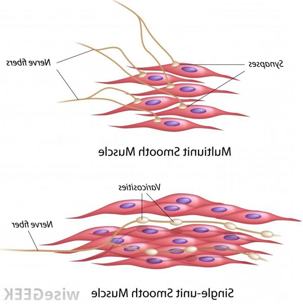
Smooth Muscle Diagram / Smooth muscle tissue. Smooth muscle tissue
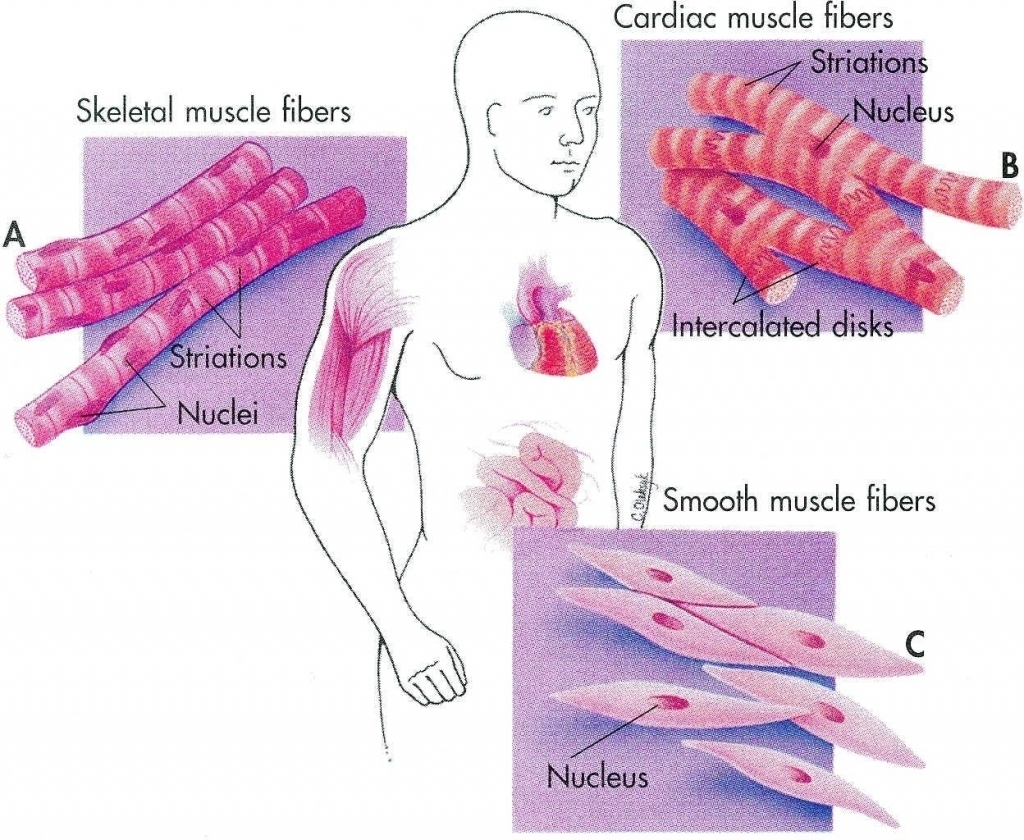
Smooth Muscle Drawing at GetDrawings Free download
Smooth Muscle Cells Are A Lot Smaller Than Cardiac Muscle Cells, And They Do Not Branch Or Connect End To End The Way Cardiac Cells Do.
View The Slide On An Appropriate Objective.
These Cells Have Fibers Of Actin And Myosin Which Run Through The Cell And Are Supported By A Framework Of Other Proteins.
In The Histology Of Hollow Organs Like The Esophagus, Intestine, You Will Find Two Concentric Layers Of Smooth Muscles In The Same Section Of That Particular Tissue.
Related Post: