Synovial Joint Drawing
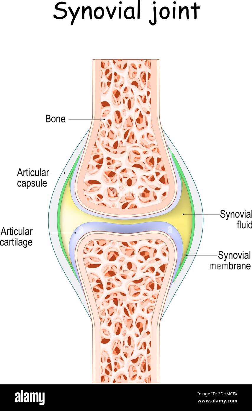
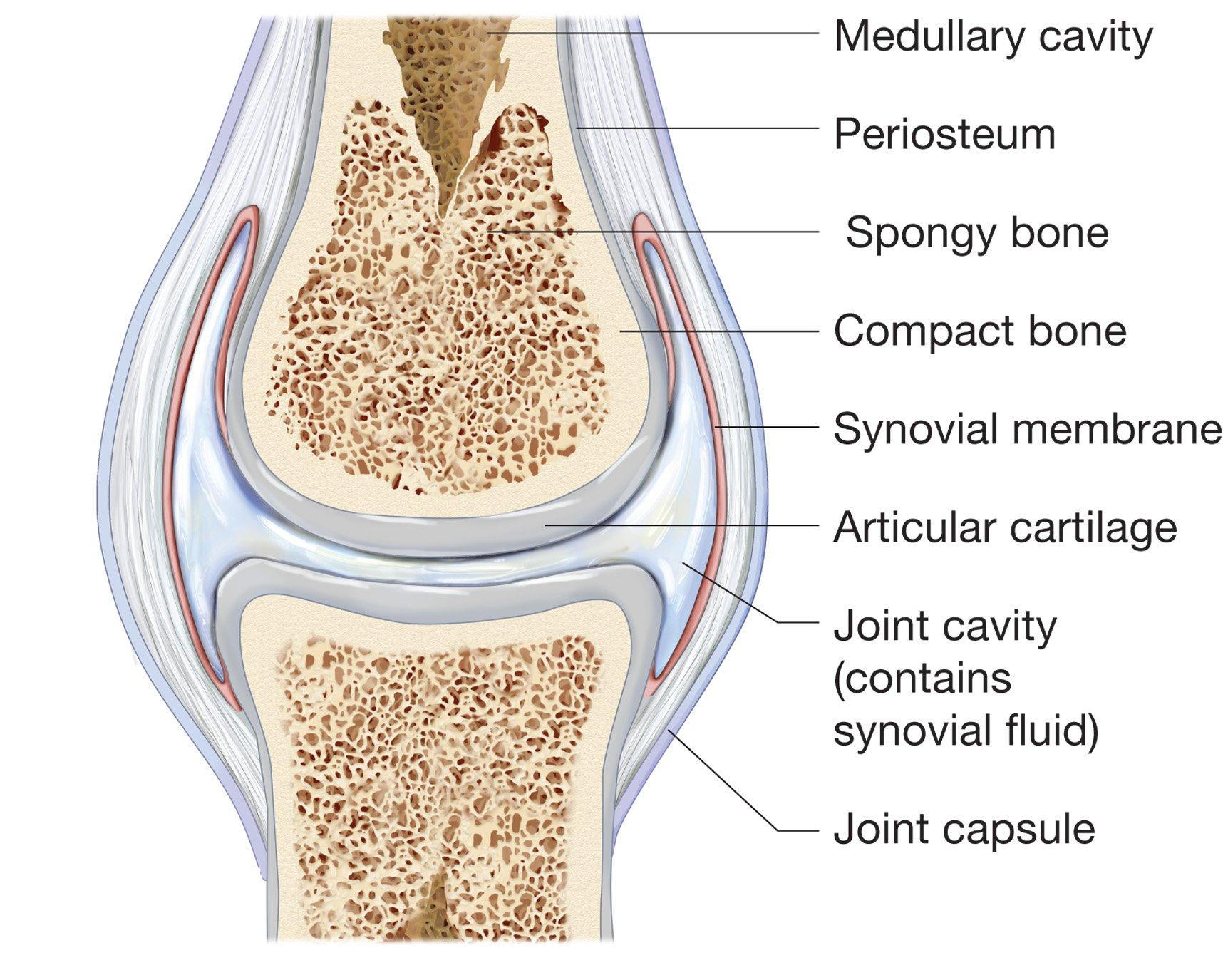
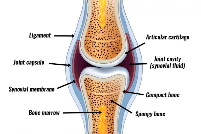
Synovial Joint Drawing - Web synovial joints are subdivided based on the shapes of the articulating surfaces of the bones that form each joint. The movement that separates a limb or other part from the axis, or middle line, of the body. These joints are termed diarthroses, meaning they are freely mobile. A key structural characteristic for a synovial joint that is not seen at fibrous or cartilaginous joints is. Diarthrosis joints are the most flexible type of joint between bones, because the bones are not physically connected and can move more freely in relation to each other. The skeletal system has a number of different joint types, for example there are fibrous joints and there are cartilaginous joints. Web synovial joints are further classified into six different categories on the basis of the shape and structure of the joint. Synovial joints are the most common type of joint in the body (figure 8.5. Web a synovial joint, also known as diarthrosis, joins bones or cartilage with a fibrous joint capsule that is continuous with the periosteum of the joined bones, constitutes the outer boundary of a synovial cavity, and surrounds the bones' articulating surfaces. (b) the hinge joint of the elbow works like a door hinge. Anatomical names for most joints are derived from the names of the bones that articulate at that joint, although some joints, such as the elbow, hip, and knee joints are exceptions to this general naming scheme. Web the joints between most of the vertebrae in the spine are cartilaginous joints. Web synovial joints are further classified into six different categories. Web this section will examine the anatomy of selected synovial joints of the body. List the six types of synovial joints and give an example of each. Web the structure and function of synovial joints is our second dash point under the skeletal system. Web a synovial joint, also known as diarthrosis, joins bones or cartilage with a fibrous joint. Web list the six types of synovial joints and give an example of each. Anatomical names for most joints are derived from the names of the bones that articulate at that joint, although some joints, such as the elbow, hip, and knee joints are exceptions to this general naming scheme. The bones of a synovial joint are surrounded by a. Web about press copyright contact us creators advertise developers terms privacy policy & safety how youtube works test new features nfl sunday ticket press copyright. You are allowed to ignore this though, as you only need to know about the synovial joints, which […] Different types of joints allow different types of movement. [1] a key structural characteristic for a. Web this section will examine the anatomy of selected synovial joints of the body. The shape of the joint affects the type of movement permitted by the joint. Also known as a diarthrosis, the most common and most movable type of joint in the body of a mammal. Web a synovial joint, also known as diarthrosis, joins bones or cartilage. Web the six types of synovial joints allow the body to move in a variety of ways. Web types of synovial joints. The following descriptions are in ascending order of mobility: You can see a drawing of a typical synovial joint in figure 14.6.2 14.6. The act of bending a joint. The bones of a synovial joint are surrounded by a synovial capsule, which secretes synovial fluid to lubricate and nourish the joint while acting as a shock absorber. Two synovial joint types are responsible for a huge range of sporting techniques involving the arms and the legs. (b) the hinge joint of the elbow works like a door hinge. Web. Web a revision lesson on how to draw and label synovial joints. Web list the six types of synovial joints and give an example of each. This joint unites long bones and permits free bone movement and greater mobility. Web about press copyright contact us creators advertise developers terms privacy policy & safety how youtube works test new features nfl. A key structural characteristic for a synovial joint that is not seen at fibrous or cartilaginous joints is. Also known as a diarthrosis, the most common and most movable type of joint in the body of a mammal. Web describe the structural features of a synovial joint. Web synovial joints are the most common type of joint in the body. The joint is surrounded by an articular capsule that defines a joint cavity filled with synovial fluid. Including what synovial fluid is, where the synovial membrane is, what joint capsules are, the ro. Web the structure and function of synovial joints is our second dash point under the skeletal system. Web a synovial joint, also known as diarthrosis, joins bones. Web describe the structural features of a synovial joint. Including what synovial fluid is, where the synovial membrane is, what joint capsules are, the ro. Planar joints planar joints have bones with articulating surfaces that are flat or slightly curved faces. Diarthrosis joints are the most flexible type of joint between bones, because the bones are not physically connected and can move more freely in relation to each other. List the six types of synovial joints and give an example of each. Synovial joints are the most common type of joint in. The joint is surrounded by an articular capsule that defines a joint cavity filled with synovial fluid. The act of bending a joint. Two synovial joint types are responsible for a huge range of sporting techniques involving the arms and the legs. The articulating surfaces of the bones are covered by a thin layer of articular cartilage. [1] a key structural characteristic for a synovial joint that is not seen at fibrous or cartilaginous joints. Web the structure and function of synovial joints is our second dash point under the skeletal system. Synovial joints are the most common type of joint in the body (figure 8.5. The following descriptions are in ascending order of mobility: The act of bending a joint. This joint unites long bones and permits free bone movement and greater mobility.
Synovial joint anatomy. joint capsule with synovial fluid and membrane

Synovial JointClassification, Definition & Examples » How To Relief

Structure and function of synovial joints HSC PDHPE
Vector Drawing Of A Synovial Joint Stock Illustration Download Image

How to draw a synovial joint easily YouTube

Synovial Joints Anatomy and Physiology I

Synovial Joint Structure

Human synovial joint Download Scientific Diagram

Structures of a Synovial Joint Capsule Ligaments TeachMeAnatomy

Normal Synovial Joint Anatomy Stock Vector Illustration of graphic
The Movement That Separates A Limb Or Other Part From The Axis, Or Middle Line, Of The Body.
Web A Revision Lesson On How To Draw And Label Synovial Joints.
You Can See A Drawing Of A Typical Synovial Joint In Figure 14.6.2 14.6.
Web 1 Key Structures Of A Synovial Joint.
Related Post:
