Transitional Epithelium Drawing
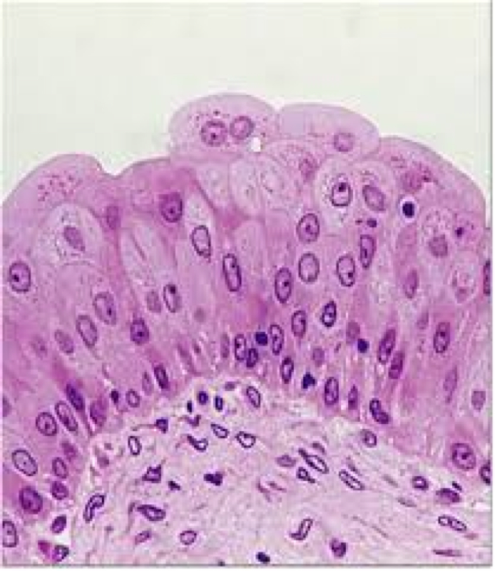
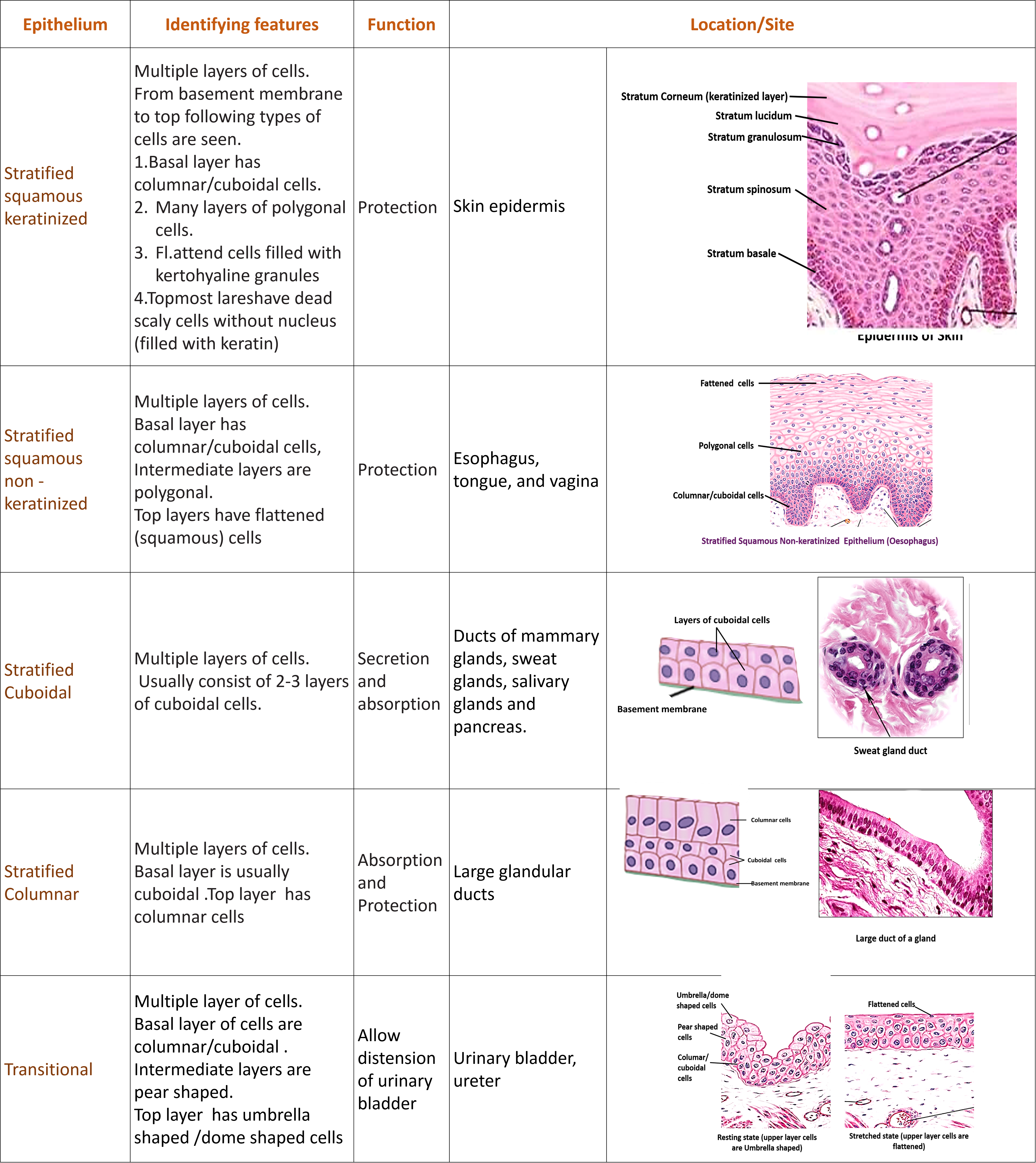
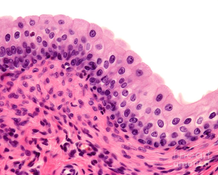
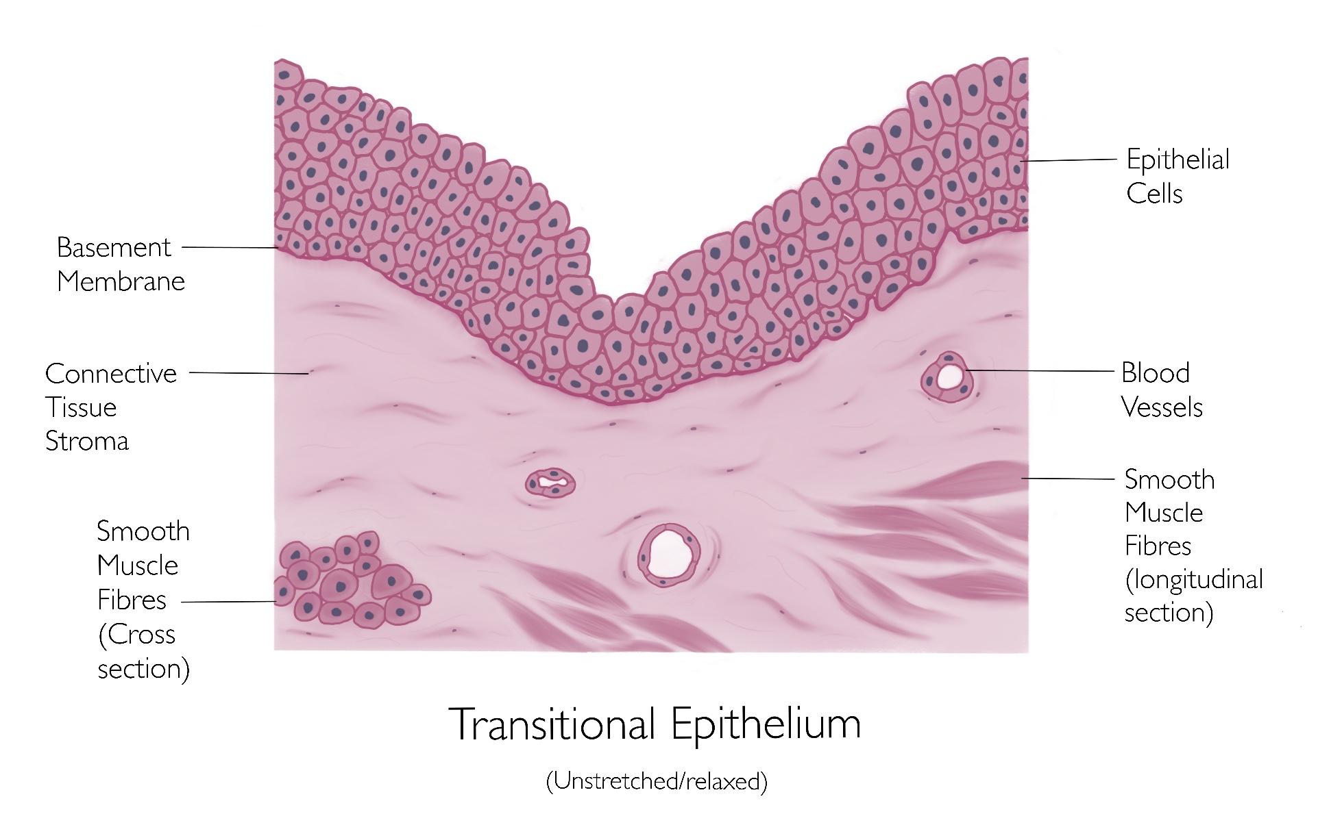
Transitional Epithelium Drawing - It is found only in the urinary system, specifically the ureters and urinary bladder. It is found only in the urinary system, specifically the ureters and urinary bladder. Web learn to draw transitional epithelium histology diagram ( for medical students) Web transitional epithelium is a stratified tissue in which the cells are all have a fairly round shape when the organ it lines is not distended (stretched out). Read more about modified simple columnar epithelium,. Web transitional epithelium animation, highlighting the epithelial layer, then underlying connective tissue. Use the image slider below to learn more about the characteristics of transitional epithelium. Web epithelial tissues provide the body’s first line of protection from physical, chemical, and biological damage. It is found only in the urinary system, specifically the ureters and urinary bladder. The epithelium has a varying appearance as they appear cubical or round when in a relaxed state, except the apical layer which. Web transcript author dan washmuth view bio instructor sarah phenix explore transitional epithelium. Read more about transitional epithelium, ureter, 40x; Web transitional epithelium definition. Web transitional epithelium is a stratified tissue made of multiple cell layers, where the cells constituting the tissue can change shape depending on the distention in the organ. Web i have described how to make transitional. Web transitional epithelium is a stratified tissue in which the cells are all have a fairly round shape when the organ it lines is not distended (stretched out). 4 views 13 minutes ago andhra pradesh. Web we will make it easy for medical students to make histology diagrams in short time during practical exams. Web transitional epithelium animation, highlighting the. Web transitional epithelium (400x) the light blue bracket indicates the transitional epithelium when the bladder is distended (full). Web transitional epithelium is a stratified tissue made of multiple cell layers, where the cells constituting the tissue can change shape depending on the distention in the organ. The epithelium has a varying appearance as they appear cubical or round when in. Contrast the messy appearance of the epithelial surface to other epithelial tissues. Web transitional epithelium is a stratified tissue in which the cells are all have a fairly round shape when the organ it lines is not distended (stretched out). Learn the definition of transitional epithelial tissue and understand its characteristics. Web i have described how to make transitional epithelium.this. When the organ is filled with fluid, cells on the topmost layer of this epithelium can stretch and appear flattened. Web we will make it easy for medical students to make histology diagrams in short time during practical exams. Web transitional epithelium is well adapted to lining the urinary tract as it can tolerate repeated stretching and recoiling (filling and. Web we will make it easy for medical students to make histology diagrams in short time during practical exams. Web transitional epithelium definition. It is found only in the urinary system, specifically the ureters and urinary bladder. It is found only in the urinary system, specifically the ureters and urinary bladder. The image shows the wall of the urinary bladder. Transitional epithelium is a type of stratified epithelium consisting of multiple layers of cells where the shape of the cell changes according to the function of the organ. Web transitional epithelium is well adapted to lining the urinary tract as it can tolerate repeated stretching and recoiling (filling and emptying with urine) without damage. 4 views 13 minutes ago andhra. Contrast the messy appearance of the epithelial surface to other epithelial tissues. Web transitional epithelial cells are epithelial cells specialized to change shape if they are stretched laterally. 4 views 13 minutes ago andhra pradesh. Transitional epithelium is a type of stratified epithelium consisting of multiple layers of cells where the shape of the cell changes according to the function. Web transitional epithelial cells are epithelial cells specialized to change shape if they are stretched laterally. The cells of an epithelium act as gatekeepers of the body, controlling permeability by allowing selective transfer of materials across its surface. The epithelium has a varying appearance as they appear cubical or round when in a relaxed state, except the apical layer which.. Web transitional epithelium (400x) the light blue bracket indicates the transitional epithelium when the bladder is distended (full). Web drawing histological diagram of transitional epithelia.useful for all medical students.drawn by using h & e pencils.explanation on epithelia while drawing.al. Transitional epithelium is a type of stratified epithelium consisting of multiple layers of cells where the shape of the cell changes. Web transitional epithelial cells are epithelial cells specialized to change shape if they are stretched laterally. The image shows the wall of the urinary bladder in the relaxed state (not distended). Web epithelial tissues provide the body’s first line of protection from physical, chemical, and biological damage. Web drawing histological diagram of transitional epithelia.useful for all medical students.drawn by using h & e pencils.explanation on epithelia while drawing.al. 4 views 13 minutes ago andhra pradesh. All substances that enter the body must cross an epithelium. Simple squamous epithelium stratified squamous epithelium simple cuboidal epithelium pseudostratified squamous epithelium simple columnar epithelium transitional epithelium Web transitional epithelium is a stratified tissue in which the cells are all have a fairly round shape when the organ it lines is not distended (stretched out). Web learn to draw transitional epithelium histology diagram ( for medical students) The cells of an epithelium act as gatekeepers of the body, controlling permeability by allowing selective transfer of materials across its surface. Web transitional epithelium animation, highlighting the epithelial layer, then underlying connective tissue. It is found only in the urinary system, specifically the ureters and urinary bladder. It is found only in the urinary system, specifically the ureters and urinary bladder. Transitional epithelium is a stratified tissue with numerous cell layers that alter shape based on organ distention. Learn the definition of transitional epithelial tissue and understand its characteristics. Web transitional epithelium is a type of stratified epithelium composed of several layers of cells, with the morphology of cells varying depending on the function of the organ.
Functions of Transitional Epithelium Tissue Video & Lesson Transcript
Epithelial Tissues Anatomy 101

The Magic Of The Transitional Epithelium How Cells Change Shape To

Histology Image Membranous epithelium

Transitional epithelium Anatomy and physiology, Histology slides
Epithelium Anatomy QA

Transitional Epithelium Diagram Quizlet
Urinary Bladder Transitional Epithelium Photograph by Jose Calvo

transitional epithelium Diagram Quizlet
Transitional Epithelium
In A Relaxed State, The Epithelium Appears Spherical Or Cubical, Except For The Apical Layer, Which May Flatten When Stretched.
Transitional Epithelium Is A Type Of Stratified Epithelium Consisting Of Multiple Layers Of Cells Where The Shape Of The Cell Changes According To The Function Of The Organ.
The Top Layer Of This Epithelium.
Web Transitional Epithelium (400X) The Light Blue Bracket Indicates The Transitional Epithelium When The Bladder Is Distended (Full).
Related Post: